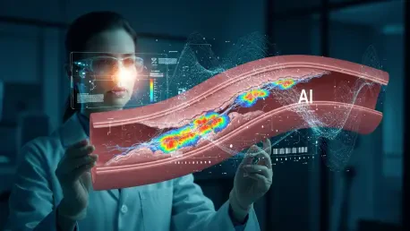

A groundbreaking artificial intelligence method offers a profound new capability in cardiovascular diagnostics, enabling clinicians to identify high-risk fatty deposits within coronary arteries with unprecedented accuracy. This innovative system, developed by a dedicated research team from the Korea Advanced Institute of Science and Technology, leverages the spectral information within existing Optical Coherence Tomography (OCT) scans to reveal what was previously invisible: the biochemical composition of arterial plaque. By combining this deep analysis with a sophisticated deep learning model, the technology promises to move beyond simple structural imaging, providing an objective and quantifiable assessment of heart attack risk. This advancement stands to transform how dangerous plaques are detected and managed, offering a powerful new tool in the fight against coronary artery disease by turning a standard diagnostic procedure into a predictive powerhouse.
Overcoming the Limits of Conventional Imaging
The primary clinical challenge this research addresses is the inherent limitation of conventional OCT technology. While OCT is a widely used and powerful tool during cardiac procedures, providing highly detailed images of a blood vessel’s interior structure, these standard images do not reveal the biochemical composition of the vessel wall. This represents a critical gap in diagnostic capability, as the makeup of atherosclerotic plaques, rather than just their size, is a key determinant of their stability. Plaques that are rich in lipids are particularly unstable and strongly associated with a higher risk of rupturing, which can trigger a blood clot and lead to a heart attack. Currently, the interpretation of OCT images to identify these high-risk plaques is a subjective process that relies heavily on the individual physician’s experience, leading to potential variability in diagnosis and risk assessment across different clinical settings.
This new method overcomes the shortcomings of traditional imaging by unlocking and analyzing spectral information that is present but underutilized in the standard OCT signal. The fundamental principle is that different types of tissue within the artery wall—such as lipid, fibrous tissue, and calcium—interact with light in unique ways. Each material absorbs and reflects light differently across various wavelengths, creating a distinct “optical signature.” The researchers’ technique successfully extracts this wavelength-dependent data from the OCT images and feeds it into a custom-developed AI model. This deep learning network is specifically trained to recognize the subtle signal patterns and spectral signatures that are characteristic of lipid-rich tissue. Consequently, the AI can automatically identify and highlight suspicious regions within the vessel wall that are likely to contain these dangerous fatty deposits, providing a quantitative and objective assessment.
A Practical and Validated Approach
A particularly innovative and practical aspect of this AI-driven system is its “weakly supervised” learning approach, which circumvents a major bottleneck in medical AI development. Many conventional AI models used in medical imaging require an incredibly labor-intensive training process where human experts must painstakingly label the exact location of the target tissue at the pixel level on thousands of images. This process is not only extremely time-consuming but can also be subjective. In contrast, the model developed by the Korean research team learns from much simpler, frame-level annotations. During the training phase, the model only needs to be told whether a given OCT image frame contains lipids or not, without specifying their precise location. This significantly reduces the annotation burden on medical experts, making the development and refinement of the AI model far more practical and scalable for real-world clinical use.
To validate the efficacy and accuracy of their method, the researchers conducted a rigorous study using intravascular imaging data obtained from a rabbit model of atherosclerosis. They meticulously compared the AI model’s predictions of lipid presence and distribution against the definitive results from histopathology, which involves physically staining tissue samples to confirm the location of lipids. The validation results were highly encouraging, demonstrating that the AI model had a strong classification performance in accurately identifying image frames containing lipid-rich plaques. Furthermore, the AI-generated heatmaps showing the location of lipids exhibited good spatial agreement with the pathological findings. This confirmed that the model was not only detecting the presence of dangerous lipids but was also highlighting anatomically meaningful and correct regions within the arteries.
Paving the Way for a New Standard of Care
The potential clinical implications of this technology were profound, as its software-based nature allowed for seamless integration into existing OCT systems found in catheterization labs worldwide without any need for hardware modifications. During a coronary intervention, this tool provided clinicians with real-time, objective information about plaque composition, which supported more informed risk assessment and aided in procedural planning, such as determining the optimal placement for a stent. This additional data allowed for a more accurate evaluation of treatment response, fostering safer clinical decision-making and enabling more individualized treatment strategies for patients with coronary artery disease. The technology ultimately had the potential to significantly improve long-term patient management and outcomes by making precision medicine a reality in interventional cardiology.
Moving forward, the research team focused on refining the technology for clinical prime time, with immediate goals that included improving the processing speed and overall robustness of the algorithm to ensure it could operate effectively in a real-time clinical setting. The plan involved conducting more extensive validation studies using data from human coronary arteries to confirm its performance in patients. A key part of future work also involved determining the most effective way to integrate the method’s output into existing clinical workflows to ensure it was intuitive and seamless for physicians to use. The underlying framework—using AI to analyze subtle signal variations in imaging data—also opened up new avenues for advanced medical diagnostics by being potentially extendable to other intravascular or optical imaging modalities.


