
The next breakthrough in cardiovascular medicine might not originate from a high-tech laboratory or a sprawling clinical trial, but from the silent, detailed images captured by an MRI machine. A revolutionary approach using artificial intelligence is now deciphering the complex visual data within heart scans, uncovering unexpected connections between existing substances and cardiac health. This technology suggests that the key to future treatments has been hidden in plain sight all along, waiting for a system intelligent enough to see it. This fusion of medical imaging and AI is fundamentally changing how researchers identify therapeutic targets, accelerating the path from discovery to potential patient care.
Unlocking New Insights from Medical Scans
For years, the search for novel heart disease therapies has followed a conventional path, relying on lengthy and costly biological experiments. However, a pioneering AI model has disrupted this process by analyzing the physical characteristics of the heart itself. By learning to recognize subtle patterns in cardiac structure and function, the system can predict which genes and drugs are most likely to influence heart conditions. This innovative method led to the counterintuitive finding that common substances, including caffeine, could exert protective effects against certain heart rhythm disorders. Such discoveries, generated not through traditional trials but through advanced computational analysis, highlight a paradigm shift in medical research where data-rich images become a primary source of biological insight.
This new frontier moves beyond simply diagnosing conditions from a scan. The AI integrates visual information with vast biological databases to create a holistic view of heart disease. It connects the dots between how a heart physically appears and the underlying genetic and molecular pathways driving its condition. This capability allows the system to generate new, testable hypotheses about disease mechanisms and potential treatments, offering a powerful tool to guide laboratory research and clinical development in more promising directions.
The Missing Link in Cardiovascular Research
The traditional process of discovering new treatments for cardiac conditions is notoriously slow and complex, often taking over a decade and costing billions. A major hurdle is identifying the right biological targets to pursue. To streamline this, scientists have developed knowledge graphs—powerful digital networks that map the intricate relationships between genes, proteins, diseases, and existing drugs. These graphs serve as comprehensive encyclopedias of biological information, allowing researchers to explore known connections and infer new ones.
Despite their utility, these knowledge graphs have always had a critical blind spot. They contained extensive information about molecular interactions and genetic predispositions but lacked data on the physical manifestation of a disease within an organ. Without information on how a condition like heart failure actually alters the shape, size, and function of the heart muscle, the predictive power of these models remained limited. This gap between abstract biological data and real-world physiology represented a significant barrier to translating genomic discoveries into effective therapies.
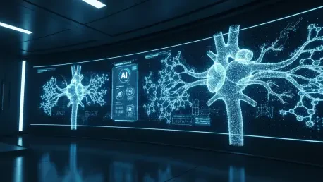
Teaching an AI to See the Heart
To bridge this gap, a research team led by Dr. Khaled Rjoob and Professor Declan O’Regan developed CardioKG, a first-of-its-kind knowledge graph enhanced with detailed cardiac imaging data. Their approach integrated high-resolution heart scans from the UK Biobank, a large-scale biomedical database. The dataset included imaging from 4,280 participants with known heart conditions, such as atrial fibrillation or heart failure, and 5,304 healthy individuals, providing a rich foundation for comparison. From these scans, the researchers generated over 200,000 unique image-based traits describing the heart’s structure and function with unprecedented detail.
This massive volume of visual data was then woven into a sophisticated network alongside information from 18 other biological databases. This comprehensive framework combined the physical characteristics of individual hearts with existing knowledge of genetics, disease pathways, and drug interactions. A powerful AI model was then trained on this integrated knowledge graph, learning to identify subtle patterns that link specific visual traits to genetic markers and potential therapeutic interventions. This process effectively taught the AI not just to recognize disease but to understand its physical signature.
From Pixels to Potential Prescriptions
The integration of imaging data yielded groundbreaking results, dramatically improving the AI’s accuracy in identifying novel genes and drugs relevant to heart conditions. By analyzing the heart’s anatomy and physiology, CardioKG pinpointed previously unknown gene-disease associations and identified promising opportunities for drug repurposing. The model suggested that methotrexate, a common treatment for rheumatoid arthritis, could be beneficial for patients with heart failure. It also proposed that gliptins, a class of drugs used for diabetes, could be repurposed to treat atrial fibrillation, a common heart rhythm disorder.
One of the most surprising discoveries was the AI’s identification of a protective effect associated with caffeine in patients with atrial fibrillation. This finding, which runs contrary to conventional wisdom that caffeine excites the heart, is supported by several other recent observational studies, adding credibility to the model’s predictive power. These results demonstrate the system’s ability to not only validate existing hypotheses but also to generate unexpected insights that can challenge long-held assumptions and open new avenues for clinical investigation.
A New Blueprint for Medical Discovery
The framework established by CardioKG serves as a powerful proof-of-concept with implications extending far beyond cardiology. This model of integrating organ-specific imaging into knowledge graphs can be adapted for virtually any medical field. For instance, neurologists could apply a similar system to brain scans to uncover new therapeutic targets for dementia, while metabolic researchers could use body-fat imaging to investigate novel obesity therapies. The potential applications are vast, promising to accelerate discovery across a wide spectrum of human diseases.
For the pharmaceutical industry, this technology offers a more rapid and efficient method for generating and prioritizing high-value biological targets for drug development. By pinpointing the most promising pathways before extensive laboratory work begins, it could significantly reduce the time and cost associated with bringing new treatments to market. The ultimate vision for this technology is to evolve it into a dynamic, patient-centered system. Such a framework could one day track an individual’s disease trajectory over time, allowing clinicians to predict illness before symptoms appear and ushering in a new era of proactive, personalized medicine. The work established a foundational blueprint that redefined how visual data could drive the future of medical innovation.


