
In the world of genetic diagnostics, some of the most critical clues are hidden in plain sight, visible only through a microscope. Yet, the human eye, for all its power, can be a subjective tool. Ivan Kairatov, a biopharma expert at the forefront of technological innovation in research and development, joins us to discuss a groundbreaking leap forward. His work centers on leveraging artificial intelligence to automate the painstaking process of counting sister chromatid exchanges—a key diagnostic marker for rare genetic disorders like Bloom syndrome. We’ll explore the limitations of the traditional manual approach, delve into the mechanics of a new machine-learning model that promises objectivity and speed, and examine how this technology performed in a crucial validation study. Finally, we’ll look toward a future where AI could become an indispensable partner in laboratories everywhere, transforming how we diagnose and understand disease at the cellular level.
Counting sister chromatid exchanges has traditionally relied on the human eye. Could you walk us through that manual process and explain where subjectivity and variability between different clinicians might arise, impacting diagnostic consistency for conditions like Bloom syndrome?
Absolutely. The manual process is incredibly labor-intensive and requires a highly trained eye. A clinician sits at a microscope, looking at specially stained chromosomes from a patient’s cells. They have to visually scan each and every chromosome, searching for these tiny, telltale segments that have been swapped between the two identical strands, or sister chromatids. It feels like a high-stakes search for a needle in a haystack. The real problem is subjectivity. What one expert perceives as a clear exchange, another might see as a smudge or an artifact of the staining process. This is where the variability comes in; two different people looking at the same slide can come up with different counts, which is deeply problematic when a high count is a strong indicator for a serious condition like Bloom syndrome, which carries a predisposition to cancer.
Your machine-learning model involves a suite of algorithms. Can you break down the distinct steps—from identifying individual chromosomes to clustering the exchanges—and explain why this multi-stage approach is more effective than a single, end-to-end analysis?
We designed it as a multi-stage pipeline because each step presents a unique, complex challenge. First, the system has to look at a raw microscope image, which is often a cluttered mess of overlapping chromosomes, and digitally separate each one. That alone is a significant image analysis task. Once a chromosome is isolated, a second algorithm takes over. Its sole job is to analyze that specific chromosome and determine whether any exchanges are present. Finally, a third algorithm aggregates the findings from all the individual chromosomes, clusters the identified exchanges, and spits out a final, objective count. A single, end-to-end model would be far less reliable. By breaking the problem down, we allow each algorithm to become a specialist at its task, leading to a much more robust and accurate overall system.
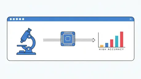
The system achieved an 84.1% accuracy rate. What does this figure represent in a practical clinical setting, and what were the biggest technical hurdles in training an algorithm to correctly identify these telltale swapped segments on stained chromosomes?
An accuracy of 84.1% is a fantastic starting point for clinical application. It means the system is reliable enough to serve as a powerful screening tool and a consistent baseline, significantly reducing the manual workload and providing a more objective measurement than what we have now. The biggest hurdle was, without a doubt, the sheer variability in the images themselves. Chromosomes don’t always lie perfectly flat, the staining isn’t always uniform, and there are countless visual artifacts that can mimic an actual exchange. We had to train the algorithm on a massive dataset to teach it the subtle visual grammar of a true sister chromatid exchange versus a shadow, a bend, or a blur. It was a painstaking process of teaching a machine to see with the discernment of a seasoned expert.
To validate the system, you analyzed cells with a suppressed BLM gene. Could you share how the algorithm’s final counts compared to those from human experts and what this consistency implies for its potential as a reliable, objective diagnostic tool?
This was the real test for us. We used cells where the BLM gene was knocked out, which artificially mimics the cellular environment of a patient with Bloom syndrome and results in a very high number of exchanges. It was the perfect high-stakes scenario. When we fed these images to our system, the counts it generated were strikingly consistent with those from our human experts. This was a watershed moment. It demonstrated that the algorithm wasn’t just accurate in theory; it could perform reliably on challenging, clinically relevant samples. This consistency is the cornerstone of a diagnostic tool. It means we have something that can deliver the same, objective result every time, removing the human subjectivity that has long been a challenge in this field.
Looking ahead, you plan to train the algorithm with more clinical data. What specific refinements or capabilities are you hoping to achieve with this larger dataset, and what are the key steps to integrating this technology into standard hospital workflows?
More data is the key to pushing this technology to the next level. By training the model on vast amounts of real-world clinical data, we expect to push that 84.1% accuracy even higher and make the system more robust against variations in lab procedures and image quality. The goal is to create a tool that is not just accurate but universally reliable. For integration, the path involves developing a user-friendly software interface that can plug directly into existing digital microscopy systems in hospitals. We’ll need to conduct further validation studies in different clinical settings to ensure its performance is consistent, and ultimately, secure the necessary regulatory approvals to make it a standard part of the diagnostic toolkit. We want to make it so simple that a lab technician can get a reliable, automated count with just a few clicks.
What is your forecast for the role of AI in automating other complex, time-consuming microscopic analyses in genetic and medical research?
I believe we are at the very beginning of a revolution. This project is a proof of concept for a much broader application of AI in medical diagnostics. Think of pathologists spending hours scanning slides for cancerous cells, or hematologists manually counting different blood cell types. Any diagnostic field that relies on a human expert identifying patterns through a microscope is ripe for this kind of AI-powered automation. In the coming years, I forecast that AI will become an indispensable partner in the lab, not to replace human experts, but to augment their abilities. It will handle the repetitive, time-consuming tasks with superhuman speed and consistency, freeing up our brilliant clinicians to focus on the most complex cases, patient interaction, and pushing the boundaries of medical science.


