
Pancreatic cancer remains one of the deadliest forms of cancer, with a disheartening 5-year survival rate lingering around just 12%, largely because it is often diagnosed at advanced stages when treatment options are limited. The scarcity of effective early detection tools worsens this challenge, leaving clinicians and patients with few ways to predict outcomes or customize therapies. However, a groundbreaking study published on August 6, 2025, in BMC Gastroenterology introduces a pioneering approach that could change this grim reality. This research unveils a sophisticated predictive model designed to assess overall survival for pancreatic cancer patients by leveraging advanced imaging and data analytics. By integrating contrast-enhanced computed tomography (CT) and magnetic resonance imaging (MRI) with clinical data, the study aims to improve prognostic accuracy and pave the way for personalized treatment plans. Led by a team of dedicated researchers, including Jiaxi Lin, this work underscores the urgent need for innovation in a field where late diagnosis often means limited hope. The model not only promises to provide deeper insights into patient outcomes but also offers a potential lifeline through risk stratification, enabling doctors to make informed decisions that could ultimately save lives. This breakthrough represents a significant step forward, addressing both the technical and clinical dimensions of a disease that has long eluded effective management strategies.
Importance of Early Detection in Pancreatic Cancer
Challenges of Late Diagnosis
Pancreatic cancer’s devastating impact largely stems from its elusive nature, often remaining undetected until it reaches advanced stages where curative options are scarce. Symptoms such as abdominal pain or jaundice typically appear late, long after the disease has progressed beyond early intervention. The absence of reliable screening tools for the general population further worsens this issue, leaving many patients diagnosed only when palliative care becomes the primary focus. This study highlights the critical need for predictive models that can identify at-risk individuals before symptoms escalate. By leveraging advanced imaging and data integration, such models could bridge the gap in early detection, offering a chance to alter the disease’s trajectory through timely action. The potential to shift diagnosis to earlier stages could mean the difference between managing a treatable condition and confronting a terminal illness.
Moreover, the anatomical challenges of pancreatic cancer add another layer of difficulty to early diagnosis. The pancreas is located deep within the abdomen, making physical exams and biopsies less feasible as routine diagnostic tools. Imaging thus becomes the cornerstone of detection, yet traditional methods often fall short in providing prognostic insights beyond confirming the presence of a tumor. The research highlights how predictive models can transform this landscape by extracting detailed data from scans to forecast survival outcomes. This approach not only addresses the diagnostic delay but also equips clinicians with actionable information to prioritize high-risk cases, potentially reducing the mortality burden through strategic intervention.
Barriers to Effective Screening
The lack of effective screening protocols for pancreatic cancer remains a significant hurdle in improving survival rates. Unlike breast or colon cancer, where mammography and colonoscopy have become standard preventive measures, no comparable tool exists for this malignancy. Current guidelines often limit screening to high-risk groups, such as those with a family history or genetic predispositions, leaving the broader population vulnerable to undetected disease progression. This gap underscores the importance of innovative approaches like the one detailed in the study, which uses multimodal data to predict outcomes even in the absence of early symptoms. Such advancements could eventually inform the development of broader screening strategies, extending the benefits of early detection to more patients.
Additionally, the financial and logistical barriers to implementing widespread screening cannot be overlooked. Advanced imaging like CT and MRI, while powerful, involves significant costs and requires specialized expertise, limiting accessibility in many healthcare settings. The study’s focus on maximizing the utility of existing imaging data through radiomics and machine learning offers a potential solution by enhancing the value derived from each scan. By refining the predictive power of these tools, the research paves the way for more efficient use of resources, potentially making early detection more feasible within constrained systems. This could mark a critical step toward democratizing access to life-saving prognostic insights for pancreatic cancer patients across diverse environments.
Role of Imaging and Radiomics in Prognosis
CT and MRI as Diagnostic Pillars
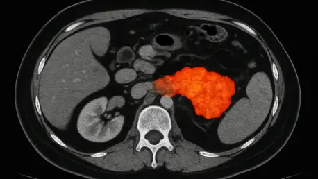
In the realm of pancreatic cancer management, imaging modalities such as CT and MRI stand as vital tools, particularly given the organ’s inaccessible location deep within the abdominal cavity. CT scans excel in delivering detailed views of tumor size, location, and potential spread to nearby structures, often revealing critical structural abnormalities like calcifications. Meanwhile, MRI provides unparalleled clarity in assessing soft tissue contrasts and vascular involvement, which are essential for understanding the tumor’s impact on surrounding anatomy. The study capitalizes on these complementary strengths by integrating data from both modalities to construct a comprehensive prognostic model. This dual approach enhances the depth of diagnostic information available to clinicians, moving beyond mere detection to offer insights into survival probabilities that can guide therapeutic strategies with greater precision.
Furthermore, the integration of CT and MRI in this predictive model addresses a longstanding limitation of relying on a single imaging type. While each modality offers unique advantages, their combined use captures a broader spectrum of tumor characteristics, from physical structure to subtle tissue interactions. The research demonstrates how this synergy can elevate prognostic assessments, providing a more nuanced understanding of disease progression. By harnessing the distinct capabilities of both imaging techniques, the model ensures that no critical detail is overlooked, enabling a more robust foundation for predicting patient outcomes. This comprehensive approach could redefine how imaging is utilized in pancreatic cancer care, setting a new standard for diagnostic thoroughness.
Radiomics: Beyond Visual Interpretation
Radiomics represents a transformative leap in medical imaging, turning subjective visual assessments into a wealth of quantitative data that can reveal hidden aspects of tumor biology. By extracting thousands of features from CT and MRI scans—such as texture, shape, and intensity—radiomics uncovers patterns that are imperceptible to the human eye, offering clues about the cancer’s aggressiveness or response potential. In this study, over 1,000 features were pulled from each imaging modality, creating a vast dataset that serves as the backbone of the predictive model. This approach shifts the paradigm from qualitative interpretation to objective analysis, providing a deeper understanding of the disease that can inform more accurate survival predictions and ultimately improve clinical decision-making.
Beyond the sheer volume of data, the application of radiomics in this research highlights its potential to bridge the gap between imaging and biological insights. Features like tumor heterogeneity or edge characteristics can correlate with underlying processes such as angiogenesis or metabolic activity, which are critical to prognosis. The study’s meticulous extraction and analysis of these elements underscore how radiomics can enhance traditional imaging by quantifying traits that influence outcomes. This innovation not only adds precision to survival forecasts but also opens the door to identifying novel biomarkers within imaging data. As a result, radiomics could become a cornerstone of personalized oncology, tailoring prognostic assessments to the unique profile of each patient’s tumor.
Multimodal Approach and Machine Learning
Combining Data for Better Accuracy
The complexity of pancreatic cancer demands a prognostic approach that mirrors its multifaceted nature, which is precisely what this study achieves by integrating multiple data sources into a single predictive model. By combining radiomic features from CT and MRI with clinical variables such as tumor marker CA125, age, and disease stage, the research constructs a holistic view of each patient’s condition. This multimodal strategy captures diverse aspects of the disease—from physical tumor characteristics to systemic indicators—resulting in more accurate survival predictions than models relying on a single data type. The synergy of imaging and clinical data reflects the intricate biology of pancreatic cancer, offering a comprehensive tool that can better inform therapeutic decisions.
Additionally, the integration of varied data types addresses the shortcomings of isolated approaches that often miss critical dimensions of the disease. For instance, while imaging alone might detail a tumor’s structure, it may not account for systemic factors like biomarker levels that influence prognosis. Conversely, clinical data without imaging might lack the spatial context needed for a full assessment. The study’s hybrid model overcomes these limitations by synthesizing these elements into a unified framework, validated by its superior performance metrics. This comprehensive method not only enhances prediction accuracy but also sets a precedent for how data integration can transform oncology, potentially extending to other complex cancers where single-modality models fall short.
Power of Random Survival Forest (RSF)
Machine learning has emerged as a game-changer in medical research, and this study leverages a specific algorithm known as Random Survival Forest (RSF) to navigate the intricacies of survival analysis. Unlike traditional statistical methods like Cox regression, which can struggle with high-dimensional data and non-linear relationships, RSF is designed to handle complex datasets with greater flexibility. It excels in managing time-to-event data, including censored observations common in cancer studies, making it ideal for predicting outcomes in pancreatic cancer. The research showcases RSF’s prowess with a C-index of 0.807, a strong indicator of its ability to distinguish between patients’ survival trajectories, surpassing other tested methods.
The superiority of RSF in this context lies in its capacity to uncover subtle patterns within vast datasets, a critical advantage when dealing with over 1,000 radiomic features alongside clinical variables. By constructing numerous decision trees and aggregating their predictions, RSF mitigates the risk of overfitting while capturing intricate interactions between variables. The study’s findings, including a Brier score of 0.101 for calibration, further affirm that RSF’s predictions align closely with actual outcomes, fostering trust in its clinical applicability. This technical edge positions RSF as a powerful tool for advancing prognostic models, potentially reshaping how survival analysis is conducted in oncology by prioritizing precision and adaptability over conventional approaches.
Clinical Impact of Risk Stratification
Guiding Treatment Decisions
One of the most practical outcomes of this predictive model is its ability to stratify pancreatic cancer patients into high- and low-risk groups based on calculated risk scores. This stratification, validated through Kaplan-Meier survival analysis, reveals stark differences in outcomes between the groups, with statistical significance confirmed by a p-value of less than 0.0001. For clinicians, this means having a reliable tool to guide treatment decisions—high-risk patients might benefit from more aggressive interventions or closer monitoring, while low-risk individuals could avoid unnecessary procedures, preserving quality of life. The model’s capacity to tailor care to individual risk profiles represents a significant advancement in personalized medicine for a disease often marked by uniform, late-stage treatments.
The real-world implications of risk stratification extend beyond individual patient care to broader healthcare strategies. By identifying which patients are most likely to face poor outcomes, resources can be allocated more effectively, ensuring that intensive therapies are directed where they are most needed. The study’s emphasis on actionable insights highlights how predictive models can bridge the gap between data and decision-making, transforming raw numbers into meaningful clinical guidance. This approach not only optimizes treatment plans but also fosters a more nuanced understanding of disease progression, enabling healthcare providers to anticipate challenges and adjust strategies proactively in the complex landscape of pancreatic cancer management.
Key Prognostic Factors
Among the numerous variables analyzed in the study, certain factors stood out as critical predictors of survival, offering valuable insights for clinical practice. The tumor marker CA125 emerged as the most influential, aligning with existing research that links elevated levels to worse outcomes in pancreatic cancer. Disease stage followed as another pivotal factor, reinforcing the well-established correlation between advanced stages and reduced survival. Additionally, specific CT-derived radiomic features contributed significantly to the model’s predictions, uncovering hidden patterns that reflect tumor behavior. These findings provide a roadmap for clinicians to focus on key indicators when assessing prognosis, blending traditional biomarkers with cutting-edge imaging data.
The identification of these prognostic factors also underscores the value of a multimodal approach in capturing the full spectrum of influences on survival. While CA125 and disease stage are familiar to oncologists, the integration of radiomic features introduces a novel dimension that enhances precision. The study’s ability to quantify the relative importance of each factor through advanced analytics like SurvSHAP(t) plots ensures that clinical attention is directed to the most impactful elements. This prioritized understanding can refine risk assessments, helping to tailor interventions more effectively. Moreover, it sets the stage for future research to explore additional variables or refine existing ones, continually improving the accuracy and relevance of prognostic tools in pancreatic cancer care.
Future Directions for Enhanced Models
Looking ahead, the study acknowledges limitations that pave the way for further refinement of predictive models in pancreatic cancer. Its retrospective design and potential selection bias, stemming from historical data over a decade, highlight the need for prospective studies to validate findings in real-time settings. Expanding research to multiple centers could also ensure the model’s applicability across diverse patient populations and healthcare systems. Such steps are crucial to confirm the robustness of risk stratification and address gaps like incomplete treatment or pathological data, which were noted as constraints in the current work. These efforts could solidify the model’s transition from research to routine clinical use.
Another promising avenue lies in incorporating advanced techniques like deep learning, which could handle even larger datasets and uncover more intricate patterns within imaging and clinical data. The study’s authors also suggest integrating additional variables, such as genetic or molecular markers, to create a truly comprehensive prognostic tool. This evolution toward richer data integration could further personalize treatment, matching risk profiles to specific therapies with greater accuracy. By addressing these future considerations, the field can build on the current model’s success, pushing the boundaries of what’s possible in pancreatic cancer prognosis and offering renewed hope for improved patient outcomes through tailored medical strategies.


