
Cardiovascular disease remains one of the leading health concerns, with aortic stenosis standing out as a particularly threatening condition. Characterized by the narrowing and stiffening of the aortic valve, aortic stenosis can lead to severe complications if not diagnosed and treated promptly. Historically, echocardiography, a type of ultrasound, has been the go-to method for diagnosing this condition. However, recent advancements have introduced four-dimensional flow MRI (4D Flow MRI) as a revolutionary tool, promising greater precision and accuracy. With widespread implications for patient care, this novel diagnostic technology has captured the attention of cardiologists worldwide.
Aortic Stenosis: A Common Yet Dangerous Condition
Symptoms and Prevalence
Aortic stenosis primarily affects older adults, with an estimated 5% of seniors over 65 in the United States experiencing its symptoms. Common signs include chest pains, rapid heartbeats, dizziness, shortness of breath, and fatigue, often mistaken as normal aging. Such symptoms can lead to misdiagnosis or delayed treatment, heightening the risk of severe complications, including heart failure. In the United Kingdom, approximately 300,000 individuals grapple with this condition, underscoring the need for accurate diagnostic tools to identify and manage these cases effectively. As the global population ages, early and precise diagnosis is crucial for mitigating the impact of this potentially fatal heart issue.
Conventional Diagnostic Methods
Traditionally, doctors have relied on echocardiography to diagnose aortic stenosis. While this method has served its purpose, its limitations are increasingly apparent. Echocardiography primarily measures blood flow velocity and valve abnormalities, presenting challenges in visualizing the complex blood flow patterns within the heart. As a result, echocardiography can produce inaccurate assessments, particularly in borderline cases where the condition’s severity is uncertain. These limitations have prompted researchers to explore more advanced imaging techniques, leading to the development and implementation of 4D Flow MRI. This technology offers an innovative solution, providing doctors with actionable insights to improve patient outcomes.
Revolutionizing Cardiological Assessments
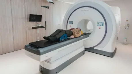
Employing 4D Flow MRI
4D Flow MRI stands as a groundbreaking method that provides three-dimensional imaging coupled with the dimension of time, allowing cardiologists to observe blood flow dynamics within the heart. By capturing a broader range of data, this advanced imaging technique furnishes detailed, precise measurements that were previously unattainable. Its capability to illustrate the flow of blood through the heart encompasses the dimensions of space and time, offering vital insights into potential surgical interventions. Developed by leading scientists at the University of East Anglia, this method has shown remarkable reliability in clinical trials, surpassing traditional diagnostic approaches’ efficacy.
Study and Results
A study published in Open Heart investigated this revolutionary technology’s performance, comparing it to standard ultrasound methods. Involving 30 patients diagnosed using both techniques, the study revealed that 4D Flow MRI provided significantly more accurate measurements. Consequently, doctors could predict the need for surgical interventions with higher precision. Over the span of eight months, researchers validated this MRI-based approach against clinical outcomes, confirming its promise in transforming diagnostic protocols. This development could herald timely treatments, effectively reducing complications and saving countless lives. Supporting institutions across the globe have echoed its potential, emphasizing its role in advancing cardiological assessment standards.
Implications and Future Prospects
Transforming Patient Care
The implications of 4D Flow MRI extend beyond improved diagnostics, suggesting profound shifts in cardiological practices. With enhanced accuracy in determining the severity of aortic stenosis, medical professionals could strategically plan interventions, minimizing patient risks associated with late-stage surgical procedures. This reduction in uncertainty fosters confidence in treatment plans, ensuring proactive management of the condition before complications arise. Furthermore, the impact of adopting advanced methodologies is mirrored in better patient outcomes, reducing healthcare costs linked to prolonged or ineffective treatments. Such benefits are pivotal as healthcare systems contend with rising demands and resource constraints.
Collaborative Developments
Cardiovascular disease remains a predominant health issue, with aortic stenosis posing a significant risk. This condition involves the narrowing and hardening of the aortic valve, potentially leading to severe health problems if not diagnosed and treated in a timely manner. Traditionally, echocardiography, a kind of ultrasound imaging, has been the primary method for identifying aortic stenosis. This non-invasive technique uses sound waves to produce images of the heart, allowing physicians to assess valve function and blood flow. However, recent technological advancements have led to the emergence of four-dimensional flow MRI (4D Flow MRI), offering enhanced precision and reliability. This cutting-edge diagnostic tool has gained attention from cardiologists globally due to its ability to provide detailed information on the heart’s functional dynamics. As these innovations unfold, they promise substantial improvements in patient care, making diagnosis more accurate and potentially improving treatment outcomes.


