
The perplexing reality that a medication meticulously designed to target cancer cells in the lungs can unexpectedly trigger adverse reactions in the heart has long been one of pharmacology’s most frustrating and dangerous puzzles. For decades, the path a drug takes after being administered has been largely obscured, creating a “black box” that conceals the precise cellular interactions responsible for both healing and harm. This fundamental lack of visibility has not only limited the effectiveness of treatments but has also been a primary driver of costly clinical trial failures and unforeseen patient side effects. Now, a revolutionary imaging technique is finally illuminating this hidden world, offering a detailed, body-wide map that promises to change how medicines are developed and understood.
The Hidden Journey of a Pharmaceutical
A fundamental question has long plagued drug development: why does a therapy intended for one part of the body cause unintended consequences in another? The answer lies in the complex and largely unobserved journey a drug takes through a living system. Once a pill is swallowed or a medication is injected, its molecules travel throughout the body, interacting with countless cells. The inability to track this journey at a granular level has been a persistent roadblock, leaving researchers to infer a drug’s behavior from indirect evidence rather than direct observation.
This “black box” problem stems from the limitations of traditional tracking methods. Techniques like radioactive imaging can provide a blurry, organ-level snapshot, confirming that a drug has reached the liver or the brain. However, they cannot reveal which specific types of cells within those complex organs the drug is actually binding to. This is the critical missing piece of the puzzle, as an organ contains a vast ecosystem of different cells, and a drug binding to one type might be therapeutic while binding to another could be toxic. The need for single-cell precision has been the holy grail of pharmacology.
A High Resolution Map for Drug Discovery
A groundbreaking imaging technique developed at Scripps Research, known as vCATCH, has emerged to solve this long-standing mystery. This technology creates an unprecedented, single-cell resolution map of a drug’s binding sites throughout an entire mammal’s body, effectively opening the black box. It moves beyond the surface-level imaging of previous methods, allowing scientists to see deep inside tissues and chart the precise cellular destinations of a given pharmaceutical.
The methodology behind vCATCH is an elegant fusion of chemistry and biology. The process begins by modifying a class of drugs known as covalent drugs—which form strong, permanent bonds with their targets—by attaching a small, inert chemical “handle.” Once this modified drug is administered and binds to cells, researchers use “click chemistry,” a Nobel Prize-winning set of reactions, to “snap” a fluorescent tag onto the handle. This illuminates every single molecule of the drug, no matter where it is in the body.
During development, the team overcame a significant hurdle where a “copper soaking” effect prevented the fluorescent signal from penetrating deep into organ tissues. Their innovative solution involves a multi-step process that first saturates the tissue with copper to block non-specific binding, followed by up to eight cycles of treatment with the fluorescent tag and catalyst. The exceptional specificity of the click chemistry reaction ensures that despite these repeated treatments, the fluorescent signal accumulates only at the intended drug sites, resulting in a clear and accurate map of cellular interactions deep within the body.
From Terabytes of Data to Cellular Targets
The sheer volume of information generated by vCATCH is immense, with a single mouse scan producing multiple terabytes of high-resolution data. To process this deluge of visual information, researchers collaborated with engineers to develop sophisticated artificial intelligence pipelines. These AI systems automatically analyze the images, identifying and mapping every single drug-bound cell across the entire organism, turning raw data into an actionable atlas of drug activity.
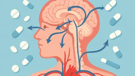
To validate the method’s accuracy and power, the team applied it to two major cancer drugs. First, they mapped afatinib (Gilotrif), a treatment for non-small cell lung cancer. The results confirmed expectations, showing the drug binding widely within lung tissue, proving the technique’s reliability. The second case study on ibrutinib (Imbruvica), a drug for blood cancers, yielded a far more significant discovery. The vCATCH map revealed a direct molecular explanation for ibrutinib’s known cardiovascular side effects, such as irregular heartbeat. It showed that the drug was binding extensively to off-target immune cells within the liver, heart tissue, and blood vessels, providing the first concrete visual evidence linking the side effects to these previously unknown cellular interactions.
Expert Perspectives on a Pharmacological Shift
This advance represents a monumental transition from ambiguity to precision. According to lead researcher Professor Li Ye, the scientific community can now move beyond the black box and directly visualize where and how drugs function in the body. The ability to see exactly which cells are affected provides a clear path to understanding not just a drug’s intended action but also its unintended consequences.
The findings related to ibrutinib are particularly impactful. They forge a direct link between a well-documented clinical side effect profile and specific, newly discovered molecular targets. This breakthrough reinforces the scientific credibility of the vCATCH approach, which builds upon the foundational Nobel Prize-winning principles of click chemistry. By providing a clear, evidence-based explanation for adverse events, the technology gives researchers the information needed to potentially redesign drugs to be more targeted and safer.
Toward a New Era of Safer Medicines
The transformative implications of vCATCH are poised to reshape the future of pharmaceutical development. The technology provides a powerful new tool for the pre-clinical screening of drug candidates. Pharmaceutical companies can now identify compounds with dangerous off-target binding profiles long before they enter costly and time-consuming human trials, significantly reducing the risk of late-stage failures and enhancing patient safety.
Beyond safety screening, the technique opens up new avenues for research into drug efficacy and other complex diseases. Scientists can now investigate whether cancer drugs are more effective at targeting tumor cells compared to healthy tissues, optimizing treatments for maximum impact with minimal collateral damage. Furthermore, the technology is being applied to neuroscience to chart the precise brain cell types bound by antidepressants and antipsychotics. This work could finally unravel the complex mechanisms behind mental health treatments, paving the way for a new generation of smarter, more effective therapies for a wide range of conditions.
The development of vCATCH provided a critical tool that transformed pharmacology from a discipline often reliant on inference into one guided by direct visual evidence. By illuminating the hidden pathways of medications at the cellular level, this technology offered an unprecedented guide for navigating the complex internal landscape of a living organism. The ability to see precisely where drugs work not only solved long-standing medical mysteries but also established a new foundation for designing the safer and more effective medicines of tomorrow.


