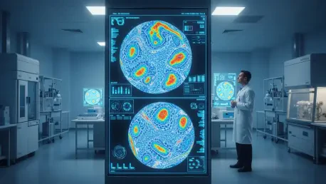

Ivan Kairatov stands at the intersection of biotechnology and digital innovation, bringing a seasoned perspective to the rapidly evolving field of computational pathology. As a veteran in biopharmaceutical research and development, he has witnessed firsthand the transition from manual microscopic observation to the current era of deep learning diagnostics. The recent findings from the University of Warwick, which analyzed over 8,000 patient samples across breast, colorectal, lung, and endometrial cancers, serve as the backdrop for this discussion. This research highlights a systemic issue where artificial intelligence often relies on statistical “shortcuts” rather than genuine biological signals, potentially compromising the integrity of patient care. In this conversation, we explore the nuances of these algorithmic biases, the narrow margin of improvement over traditional methods, and the path toward a more rigorous, causally-aware future for oncology tools.
AI models often rely on correlations, such as using microsatellite instability to predict specific gene mutations, rather than identifying direct biological signals. How do these “shortcuts” compromise diagnostic accuracy when markers do not co-occur, and what specific risks does this pose for patients in real-world clinical settings?
The fundamental danger lies in the difference between observing a symptom and understanding a cause. It is much like trying to judge the excellence of a kitchen solely by the length of the line outside; the queue is a visible shortcut, but it tells you nothing about the ingredients or the chef’s technique. In pathology, when a model learns to identify a BRAF mutation by spotting its common neighbor, microsatellite instability (MSI), it creates a statistical mirage of accuracy. The moment a patient presents with a BRAF mutation without that accompanying MSI signature, the AI fails because it never actually “saw” the mutation to begin with. In a high-stakes clinical setting, this means a patient could be denied a life-saving targeted therapy or given an ineffective treatment simply because the algorithm relied on a visual coincidence rather than the hard biological truth.
Computational tools currently achieve around 80% accuracy in biomarker prediction, which is only a modest improvement over the 75% accuracy of traditional tumor grading. Why is this performance gap so narrow, and what steps must developers take to ensure AI provides genuine information gain over standard manual assessments?
This narrow five-percent gap is a clear signal that our current models are largely just automating the intuition that pathologists have utilized for decades. When an AI hits an 80% accuracy ceiling, it is often just picking up on the same high-level tissue features—like the shape of cells or the density of the stroma—that define a standard tumor grade. To achieve a true breakthrough, we must move beyond simply building bigger models and instead implement stricter evaluation protocols that force the software to “stop cheating.” Developers need to demonstrate that their tools provide specific information gain that cannot be gleaned from a simple, pathologist-assigned grade. Until we prioritize this biological depth over headline-grabbing accuracy scores, we are merely digitizing the status quo rather than advancing the frontier of cancer diagnostics.
When diagnostic algorithms are tested within specific patient subgroups, such as those with high-grade tumors, their performance often declines sharply. How should researchers design more rigorous, bias-aware evaluation protocols to expose these dependencies, and what metrics are most essential for validating a tool’s clinical readiness?
The sharp decline in performance within stratified subgroups, such as MSI-positive tumors, reveals that many algorithms are essentially leaning on a “crutch” of confounding factors. To fix this, we need to move away from aggregate accuracy scores and embrace a more granular, bias-aware evaluation strategy that tests the model under pressure. Researchers must conduct rigorous subgroup testing where these confounding visual signals are controlled, forcing the algorithm to prove it can identify the biomarker in isolation. The most essential metric for clinical readiness isn’t just a high percentage of correct guesses, but the “information gain” the tool provides relative to a simple clinical baseline. We need to see how the model behaves when the “shortcuts” are removed, ensuring it remains reliable for the individual patient whose case might not fit the standard statistical mold.
AI is currently viewed more as a tool for clinical triaging or drug development rather than a full replacement for molecular testing. In what ways can clinicians safely integrate these tools into existing workflows today, and what specific biological relationships must future models master to become truly reliable?
Right now, these tools are best utilized as a sophisticated safety net for triaging or as a supplementary decision support system rather than a primary diagnostic engine. Clinicians can use AI to screen large volumes of slides to identify high-priority cases for molecular testing, thereby speeding up the workflow without bypassing the gold standard of laboratory verification. However, for these tools to eventually stand on their own, future models must master the causal structures of cancer biology, moving from mere correlation to a deep understanding of how specific mutations physically manifest in tissue architecture. We must be cautious not to let the excitement of “innovation” outpace the rigorous assessment of what is actually relevant and correct for a specific person. It is an imperfect first step, but we must use this period to refine our understanding of the complex, variable relationships between a tumor’s appearance and its genetic core.
Effective oncology tools must account for the immense complexity and variability of individual patient features. How can developers transition from correlation-based learning to models that explicitly grasp causal biological structures, and what are the primary technical hurdles in capturing these nuanced patterns?
The transition requires a fundamental shift in how we train these systems, moving away from simple pattern matching and toward models that explicitly incorporate biological logic and causal relationships. One of the primary technical hurdles is the sheer variability of human tissue; every patient’s cancer is a unique ecosystem, and a model trained on generalities often struggles with these nuanced, individual features. Developers must move toward grounded tailoring, where the AI is built to recognize the specific biological “why” behind a visual pattern rather than just the “what.” This means the roadmap for the next generation of pathology AI isn’t just about more data, but about better, more biologically-grounded data and evaluation standards that account for the messy reality of clinical samples. We have to embrace the complexity of the human body rather than trying to oversimplify it into a set of predictable pixel correlations.
What is your forecast for AI in cancer pathology?
My forecast is that we are entering a “correction phase” where the industry will shift its focus from raw predictive power to biological transparency and causal reliability. We will likely see a move away from the “black box” approach in favor of models that can explain their reasoning through the lens of established pathology, essentially learning to “understand meteorology” rather than just “looking for umbrellas.” Over the next few years, I expect to see the emergence of much stricter regulatory standards that mandate subgroup testing and proof of information gain before any AI tool is permitted for routine clinical use. Ultimately, AI will not replace the pathologist or the molecular test, but it will become an indispensable partner that is finally capable of deciphering the hard biology hidden within the image, leading to more precise and personalized care for every patient.


