
I’m thrilled to sit down with Ivan Kairatov, a distinguished expert in biopharma with a profound understanding of technological innovation and research in the field. Today, we’re diving into the groundbreaking advancements in biomedical imaging, specifically focusing on a pioneering project at the University of Arizona that promises to transform how we see and treat conditions like skin cancer. Ivan brings a unique perspective on how cutting-edge tech intersects with clinical needs, and we’ll explore topics ranging from innovative imaging techniques to their potential impact on patient care, the challenges of current methods, and the exciting possibilities that lie ahead in non-invasive diagnostics.
How did it feel to be part of a select group chosen for significant funding from a prestigious initiative like the NIH’s Common Fund Venture Program?
It was an incredible honor to be among just a handful of teams recognized for this initiative. The validation from such a respected body as the NIH really underscored the potential impact of our work. It felt like a huge vote of confidence in our vision to push the boundaries of imaging technology, and it motivated us even further to deliver on our goals. The excitement was paired with a sense of responsibility to make the most of this opportunity and advance healthcare solutions.
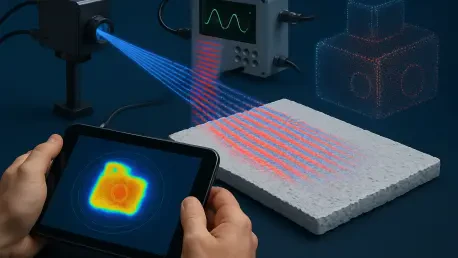
Can you walk us through the concept of synthetic wavelength imaging and why it’s such a game-changer for looking deeper into tissues?
Absolutely. Synthetic wavelength imaging, or SWI, is a novel approach where we combine two distinct illumination wavelengths to computationally create a virtual, longer wavelength. This synthetic wavelength is less affected by light scattering, which is a major hurdle when imaging deep into biological tissues. What’s exciting is that while we gain depth penetration, we still retain the high contrast from the original wavelengths. This balance allows us to see clearer, deeper images without sacrificing detail, which is a significant leap forward compared to traditional optical methods.
Why did your team choose to focus specifically on nonmelanoma skin cancers like basal cell and squamous cell carcinoma for this project?
Nonmelanoma skin cancers present unique challenges that make them a critical area of need. Unlike melanoma, which often has more distinct visual markers, these cancers vary widely in size, depth, and invasion patterns, making them harder to assess accurately with current imaging tools. They’re also incredibly common, being the most prevalent malignancies worldwide. We saw a pressing need for better, non-invasive ways to evaluate these lesions, especially to improve diagnosis and treatment monitoring for patients who often face complex care pathways.
What are some of the biggest limitations you’ve encountered with existing imaging technologies like confocal microscopy or optical coherence tomography?
Current methods like confocal microscopy and optical coherence tomography are fantastic for shallow depths, offering great resolution and contrast. However, their shorter wavelengths struggle with light scattering as you go deeper into tissue, which blurs the image and limits what you can see. On the other hand, longer wavelength options like ultrasound can penetrate deeper but often lack the detail or contrast needed for precise cancer imaging. Our goal is to bridge that gap by developing a technology that doesn’t force a tradeoff between depth and clarity.
In what ways do you see this new imaging technology transforming the diagnosis and management of skin cancer for doctors and patients?
The potential is transformative. With this technology, doctors could assess tumor margins with much greater precision right at the point of diagnosis, which is crucial for planning effective treatment. It also offers the ability to monitor how a lesion responds to therapy over time, allowing for real-time adjustments to care plans. Perhaps most importantly, by providing detailed, non-invasive imaging, we could reduce the reliance on biopsies or other invasive procedures, making the process less stressful and risky for patients while still ensuring accurate results.
Looking beyond skin cancer, how do you envision the broader applications of synthetic wavelength imaging in other areas of medicine?
The versatility of synthetic wavelength imaging opens up a wide array of possibilities. Because of its ability to tune wavelengths and penetrate scattering tissues, it could be adapted for detecting other cancers, like breast cancer, where deep tissue imaging is critical. There’s also potential for neurological applications, such as imaging inside the brain to study or diagnose conditions non-invasively. The computational aspect of SWI means we can keep refining algorithms to tackle different challenges, making it a flexible tool for many biomedical fields.
What is your forecast for the future of non-invasive imaging technologies in healthcare over the next decade?
I’m incredibly optimistic about where non-invasive imaging is headed. Over the next decade, I expect we’ll see a convergence of advanced optics, computational power, and artificial intelligence that will revolutionize diagnostics. Technologies like synthetic wavelength imaging will likely become more accessible and integrated into routine clinical practice, enabling earlier detection of diseases and personalized treatment plans. We’re also likely to see these tools become smaller and more portable, bringing high-end imaging directly to patients, even in remote or underserved areas. It’s an exciting time, and I believe we’re just at the beginning of unlocking the full potential of these innovations.


