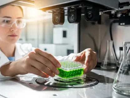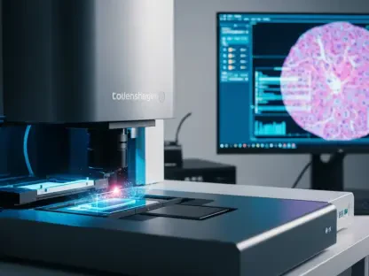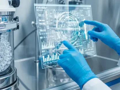

A groundbreaking advancement in medical imaging technology developed by researchers at the University of Houston promises to significantly transform medical diagnostics. This innovative development centers around a sophisticated 3D imaging system that offers enhanced precision and efficiency compared to traditional diagnostic methods. Spearheaded by Mini Das, a Moores Professor at UH’s College of Natural Sciences and Mathematics and the Cullen College of Engineering, this technology represents a monumental shift in the way medical conditions are detected and diagnosed.
The Limitations of Traditional Imaging
Conventional 2D X-rays
Historically, medical practitioners have relied on conventional 2D X-ray imaging techniques to diagnose a variety of conditions, such as bone fractures. Although 2D X-rays have been a cornerstone of diagnostic medicine for decades, their limitations have become increasingly evident. These conventional techniques often fail to detect finer details, including small fractures and soft tissue damage, which are critical in diagnosing conditions such as cancerous tissues. The inability to capture intricate details within the body poses significant challenges, particularly in early detection and accurate diagnosis.
Conventional 2D X-rays collect photons in a manner analogous to viewing white light without separating its constituent colors. This method limits their ability to distinguish between different materials beyond basic density variations. As a result, medical practitioners may struggle to identify the specific nature of an abnormality or distinguish between various types of tissue. This shortcoming necessitates the need for more advanced imaging techniques to ensure accurate and timely medical diagnoses.
MRI Scans
While MRI scans offer more detailed imaging capabilities compared to 2D X-rays, they are not without their drawbacks, being both costly and time-consuming. These factors make MRI scans less suitable for certain screening and detection environments, where rapid and efficient diagnostics are essential. Recognizing these limitations in both 2D X-rays and MRI scans, Mini Das and her team at the University of Houston embarked on a mission to develop a more effective and precise imaging solution.
MRI scans operate by using powerful magnetic fields and radio waves to produce detailed images of the body’s internal structures. Despite their high-resolution output, the financial and logistical constraints of MRI technology hinder its widespread use in routine medical diagnostics. The need for an imaging solution that balances precision, cost, and efficiency led Das and her team to explore alternative approaches, culminating in the development of an innovative 3D imaging system that promises to bridge the gap between accuracy and accessibility.
The Innovative 3D Imaging Solution
Photon-Counting Detectors
Das’s team has pioneered a novel 3D imaging solution that utilizes photon-counting detectors in conjunction with advanced algorithms. Unlike traditional X-ray technology, which captures X-rays at a single energy level, photon-counting detectors capture X-rays at multiple energy levels simultaneously. This multi-energy capture allows for a more detailed and precise visualization of different tissues and contrast agents within the body, revolutionizing the field of medical imaging.
Photon-counting detectors represent a significant departure from conventional techniques. By capturing X-rays at multiple energy levels, they provide a more nuanced understanding of the body’s internal structures. This method is akin to using a prism to split white light into its constituent colors. Just as a prism reveals the spectrum of colors within white light, photon-counting detectors reveal the specific materials within the body, enhancing the ability to differentiate between various tissues and substances. This advanced imaging capability has profound implications for improving diagnostic accuracy and patient outcomes.
Enhanced Visualization
The multi-energy capture facilitated by photon-counting detectors allows for an unparalleled level of 3D visualization. Conventional X-rays, which collect photons as a whole, can only provide a limited view, much like seeing white light without its component colors. In contrast, photon-counting detectors can distinguish between different tissues and contrast agents with remarkable precision, enabling a more comprehensive and accurate view of the body’s internal structures.
This refined visualization capability holds significant promise for medical diagnostics. For instance, materials like aluminum, plastic, iodine, or gadolinium—which are commonly used in medical imaging—can be accurately identified and quantified within the body. This ability to distinguish between various materials and tissues goes beyond basic density variations, offering a more detailed and precise analysis. Consequently, medical practitioners can make more informed decisions, improving the diagnostic process and potentially leading to better patient outcomes.
Potential for Improved Cancer Detection
Dual Contrast Agents
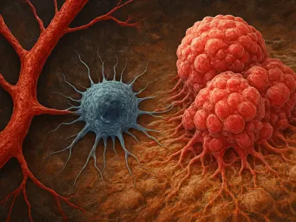
One of the most promising applications of this 3D imaging technology lies in its potential to enhance cancer detection. By injecting patients with two different contrast agents—one designed to target tumors and the other aimed at targeting inflammation—medical practitioners could more accurately pinpoint the accumulation areas of these agents. This dual-contrast approach provides a clearer distinction between malignant and benign tissues, enabling earlier and more precise cancer detection.
The ability to use two contrast agents simultaneously addresses a critical limitation of conventional imaging techniques. While traditional methods can highlight bright areas within the body, they often lack the specificity needed to determine the exact nature of these areas. The new 3D imaging technology, with its advanced photon-counting capabilities, provides a more definitive analysis, revealing not only the presence of an object but also the materials and quantities involved. This level of detail is crucial for accurately diagnosing and treating cancer, ultimately improving patient prognosis.
Quantitative Analysis
The new 3D imaging technology also offers enhanced quantitative analysis, distinguishing it significantly from current imaging methods. This capability allows for a more precise measurement of materials and tissues within the body, providing detailed information about the nature and extent of abnormalities. This detailed analysis is particularly valuable in cancer detection, where understanding the specific characteristics of a tumor is essential for developing effective treatment plans.
Quantitative analysis enables medical practitioners to assess the exact composition and distribution of tissues and materials within the body. This detailed information helps in formulating more targeted and effective treatment strategies. The ability to identify and quantify materials accurately reduces the likelihood of misdiagnosis and ensures that patients receive the most appropriate interventions. Consequently, this advanced imaging technology holds the potential to significantly improve diagnostic accuracy and patient outcomes, particularly in the field of oncology.
Overcoming Technical Challenges
Material Differentiation
Despite its promise, the new 3D imaging technology faces certain challenges that need to be addressed. One of the primary challenges is the difficulty in distinguishing between materials that exhibit similar X-ray properties. This limitation makes it challenging to differentiate more than two or three substances simultaneously within the body. Overcoming this hurdle is essential to fully realize the potential of the technology in providing detailed and accurate medical diagnoses.
To address this challenge, Das’s team is developing sophisticated methods and algorithms to improve material differentiation. By calibrating the photon-counting detectors with known materials, the team aims to correct distortions and improve the accuracy of material identification. These efforts are crucial for enhancing the reliability and precision of the technology, ensuring that it can distinguish a wider range of materials and provide more comprehensive diagnostic information.
Detector Calibration
Another significant challenge facing the development of this 3D imaging technology is the issue of detector calibration. Errors and inaccuracies in the detectors can hinder their ability to effectively separate photons by energy, compromising the quality of the imaging results. Das’s team is actively working on refining the calibration techniques to ensure that the detectors provide consistent and accurate measurements, which are critical for reliable diagnostics.
To overcome calibration challenges, the team is employing innovative approaches to calibrate the detectors with high precision. By using known materials and applying advanced algorithms, they aim to minimize errors and enhance the detectors’ performance. These efforts are vital for ensuring that the technology can deliver high-quality imaging results consistently, paving the way for its adoption in clinical settings. The successful calibration of the detectors will be a significant milestone in the development of this revolutionary 3D imaging system.
Future Prospects and Applications
Collaboration with Industry Partners
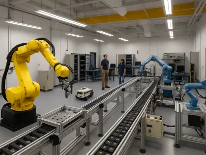
While still in the research and development stage, the project has made significant strides, thanks to collaboration with industry partners. Das’s team is working with industry experts in Europe to create larger detectors and optimize their performance. This collaboration is essential for scaling up the technology and preparing it for real-world applications. By leveraging the expertise and resources of industry partners, the team aims to bring this groundbreaking technology closer to practical use.
The current detectors are relatively small, and there is a need to enhance their measurement accuracy before they can be deployed in clinical settings. The collaboration focuses on refining the detector technology and addressing any remaining challenges. These efforts are crucial for transitioning the technology from the laboratory to real-world applications, where it can have a significant impact on medical diagnostics. The success of these collaborative endeavors will be a key factor in the widespread adoption of this advanced 3D imaging system.
Beyond Medical Imaging
The potential applications of this 3D imaging technology extend well beyond the realm of medical diagnostics. In addition to its profound impact on healthcare, the technology holds promise for various other fields, including materials science, security, geophysics, and micro- and nano-electronics imaging. The ability to capture detailed and accurate 3D images has wide-ranging implications, offering new possibilities for research and innovation in these diverse areas.
For instance, in the field of security, the technology could be used for advanced baggage scanning, enhancing the detection of potential threats. In materials science, it could enable detailed analysis of material properties and behaviors, leading to new discoveries and innovations. Similarly, in geophysics, the ability to capture precise 3D images could improve the understanding of geological structures and processes. The technology’s versatility and advanced capabilities make it a valuable tool for various applications, highlighting its potential to drive progress and innovation across multiple domains.
Support and Funding
Previous Research Contributions
Das’s prior work in exploring the wave nature of X-rays has significantly contributed to the enhancement of contrast in soft materials. This research, which was previously featured in the prestigious scientific journal Optica, laid the groundwork for the development of the current 3D imaging technology. By focusing on the fundamental properties of X-rays, Das’s team was able to pioneer new methods for improving imaging contrast and accuracy.
This foundational research provided valuable insights into the behavior of X-rays and their interactions with different materials. These insights played a crucial role in the development of the photon-counting detectors and advanced algorithms used in the new 3D imaging system. The lessons learned from this earlier research have been instrumental in overcoming the technical challenges associated with the new technology, paving the way for its successful development and implementation.
Funding Agencies
The revolutionary advancement in medical imaging technology developed by researchers at the University of Houston promises significant improvements to medical diagnostics. At the core of this groundbreaking development is an advanced 3D imaging system that provides better precision and efficiency compared to conventional diagnostic methods. Led by Mini Das, a Moores Professor at UH’s College of Natural Sciences and Mathematics and the Cullen College of Engineering, this innovation marks a substantial shift in the detection and diagnosis of medical conditions. The enhanced 3D imaging system could lead to faster, more accurate diagnoses, potentially improving patient outcomes and reducing healthcare costs. By providing clearer and more detailed images, doctors can make more informed decisions about treatment plans. This technological advancement represents a major leap forward in medical imaging, offering the potential to revolutionize the way healthcare professionals diagnose and treat various diseases.