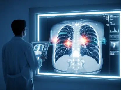

The ability to visualize the intricate cellular ballet that precedes a formal disease diagnosis has long been the primary objective of modern medicine, a goal that promises to shift healthcare from a reactive to a proactive model. At the heart of this pursuit is Positron Emission Tomography (PET), a powerful imaging technology that provides a real-time window into the body’s metabolic processes. However, a persistent chemical challenge has long constrained the full potential of PET scans, limiting the scope of diseases that can be effectively diagnosed and monitored. A recent breakthrough from a team at Virginia Tech has now shattered this long-standing barrier, heralding a new era in molecular imaging and personalized medicine.
A Glimpse into the Future of Proactive Medicine
The central promise of advanced medical diagnostics lies in the capacity to see disease not just when it manifests as symptoms, but at its nascent, cellular level. Proactive intervention depends entirely on detecting these subtle biological changes early. This is where PET imaging excels, moving beyond the anatomical snapshots of X-rays or MRIs to reveal functional information about how tissues and organs are operating. By tracking metabolic activity, PET scans can identify abnormalities long before they become structurally apparent.
Despite this capability, the application of PET has been hampered. The technology’s potential to diagnose a vast range of conditions, from neurodegenerative disorders to specific cancers, has been curtailed by a fundamental limitation in creating the very tools that make imaging possible. The gap between what PET could theoretically achieve and its practical clinical use has represented a significant hurdle in the advancement of diagnostic medicine, a hurdle that has finally been overcome.
The Unseen Bottleneck in Advanced Diagnostics
PET imaging operates by detecting signals from radioactive “tracers,” specialized molecules injected into the body that travel to specific cells or tissues. These tracers are designed to participate in metabolic processes, effectively lighting up areas of high or abnormal cellular activity. The choice of the radioactive isotope attached to the tracer is critical for clinical success.
The preferred radioisotope for this purpose is fluorine-18, largely due to its practical two-hour half-life. This duration is long enough to allow for the synthesis of the tracer, quality control, and administration to the patient, yet short enough to minimize radiation exposure. In contrast, the alternative, carbon-11, has a half-life of only about 20 minutes, severely restricting its utility. The challenge, however, has been a chemical one: attaching fluorine-18 to many of the most effective modern drug molecules has proven exceptionally difficult, particularly for those containing a trifluoromethyl group—a common component that enhances a drug’s stability and potency.
How a Copper Bridge Solved a Decades-Old Puzzle
A research team led by Virginia Tech chemist Wei Liu has developed a novel method that elegantly solves this decades-old chemistry problem. Their innovative approach uses copper as a catalyst to create a chemical “bridge,” enabling the efficient transfer of fluorine-18 to the previously inaccessible trifluoromethyl groups on complex molecules. This breakthrough effectively unlocks a vast library of compounds that can now be developed into new PET tracers.
The new technique is also remarkably versatile. Beyond its primary application with fluorine-18, the method is adaptable for use with carbon-11, offering researchers unprecedented flexibility in tracer design. This means that complex, drug-like molecules, once considered impossible to tag for PET imaging, can now be labeled and studied. This development opens the door to creating highly specific diagnostic tools that can target a multitude of biological processes with pinpoint accuracy.
A Game Changer for Molecular Imaging
The inability to label trifluoromethyl-containing molecules has been widely regarded as a major roadblock for the entire field of molecular imaging. The consensus among researchers was that this limitation severely stunted the development of new diagnostic agents, leaving many diseases beyond the reach of PET technology. Liu’s work directly addresses and resolves this fundamental barrier.
The key finding of the research is the dramatic expansion of the molecular toolkit available to scientists. By providing a reliable method to create fluorine-18 labeled tracers from a wide range of established and experimental drugs, the discovery paves the way for new avenues of scientific exploration. It allows researchers to visualize biological targets that have, until now, remained invisible, transforming the landscape of what is possible in disease detection and therapeutic development.
Charting a New Course for Disease Detection and Treatment
The immediate implication of this breakthrough is profound. Scientists can now develop a new generation of imaging agents to visualize biological targets associated with a host of diseases that are currently difficult to detect in their early stages. This capability is expected to accelerate research in fields such as oncology, neurology, and cardiology, where early and accurate diagnosis is critical for patient outcomes.
Ultimately, this advancement points toward a future defined by precision medicine. A wider array of highly specific PET tracers will not only lead to earlier diagnoses but also help guide targeted, personalized therapies. By visualizing how a specific drug interacts with its target in a particular patient, clinicians could tailor treatments for maximum efficacy and minimal side effects. The development had shifted the paradigm of medical imaging, moving the field closer to the ultimate goal of detecting and treating disease before it ever takes hold.