
Metastatic spinal tumors originating from lung cancer often cause debilitating symptoms including chronic pain, pathological fractures, and potential paralysis. In the current medical landscape of 2026, clinicians face the daunting task of managing these secondary growths with extreme precision. The spine remains the most frequent site for skeletal metastasis in lung cancer patients, often leading to a rapid decline in quality of life if not addressed through targeted intervention. Identifying the specific histological subtype, such as lung adenocarcinoma or lung squamous cell carcinoma, is no longer considered a luxury but a fundamental requirement for selecting the appropriate treatment regimen. These subtypes respond differently to chemotherapy and emerging immunotherapies, yet obtaining this information traditionally required invasive bone biopsies. Such procedures are inherently risky, especially for fragile patients whose tumors are located near the spinal cord, necessitating the development of safer alternatives. Furthermore, the anatomical complexity of the vertebral column often complicates needle placement, increasing the likelihood of procedural complications or insufficient tissue sampling. Consequently, the search for a noninvasive diagnostic tool has intensified as oncology moves toward more personalized care.
Advancements in Molecular Imaging
The Mechanism: Amide Proton Transfer-weighted Imaging
Amide Proton Transfer-weighted (APTw) imaging has emerged as a transformative development in the field of molecular magnetic resonance imaging. Unlike conventional MRI sequences that primarily map the distribution of water and fat to create anatomical images, APTw imaging utilizes a technique known as magnetization transfer. This specific method allows radiologists to detect the presence of exchangeable protons attached to amide groups in mobile proteins and peptides within the cellular environment. Because malignant cells are characterized by accelerated metabolic activity and a high rate of protein synthesis, they exhibit a distinct APTw signal that differs from healthy tissue. This provides a unique window into the biochemical makeup of a tumor, allowing for a characterization that goes beyond mere physical size or shape. By focusing on the internal chemistry of the lesion, this technology bridges the gap between traditional imaging and histopathology, offering a detailed view of cellular behavior. It represents a paradigm shift in how radiologists interpret spinal oncology, moving toward a more functional assessment.
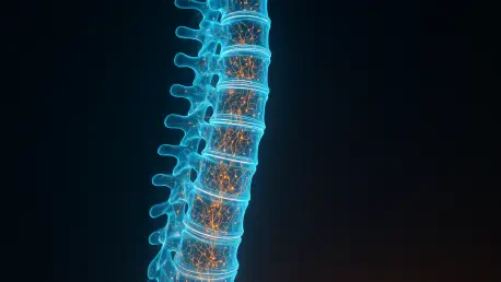
The technical execution of APTw scans involves saturating the resonance frequency of the amide protons, which then transfer their magnetization to the surrounding water molecules. This transfer results in a detectable signal intensity change that can be quantified and visualized on a heat map, representing the concentration of mobile proteins. In the context of spinal metastases, this means that clinicians can now observe the “protein density” of a tumor in real-time. This level of detail is critical because different lung cancer subtypes possess varying protein profiles. For instance, the secretory nature of certain adenocarcinomas produces a different amide proton signature than the keratin-rich structure of squamous cell carcinomas. As a result, the APTw sequence serves as a sensitive probe for the underlying metabolic processes that define a tumor’s identity. This advancement represents a shift from observing what a tumor looks like to understanding what it is actually doing at a molecular level within the spine. Such insights are proving invaluable for differentiating aggressive growths from benign vertebral changes.
Visualizing Protein Density: The Role of Mobile Peptides
One of the most significant advantages of APTw imaging is its ability to provide high-resolution metabolic data without the use of exogenous contrast agents. For decades, gadolinium-based contrast media have been the gold standard for enhancing tumor visibility in MRI; however, these agents are not without risks. Patients with impaired renal function, a common occurrence in advanced cancer cases, are often ineligible for contrast-enhanced scans due to the risk of nephrogenic systemic fibrosis. APTw imaging sidesteps this issue entirely by relying on the body’s own endogenous proteins to generate signal contrast. This “label-free” imaging approach ensures that a wider demographic of patients can receive accurate diagnostic workups without the threat of toxicity. Moreover, the absence of contrast agents simplifies the imaging workflow and reduces the overall cost and time associated with the procedure. This makes it an ideal candidate for frequent monitoring of tumor progression or response to therapy. It effectively lowers the barrier to high-quality oncological assessment in a variety of clinical settings.
Beyond safety considerations, the protein-sensitive nature of this MRI technique provides a more nuanced understanding of the tumor microenvironment than previous methods could achieve. While traditional diffusion-weighted imaging (DWI) provides information about cellular density by measuring water movement, it often fails to distinguish between different types of highly cellular tumors. APTw imaging complements these physical metrics by adding a layer of chemical information, specifically targeting the mobile peptides that are abundant in aggressive lung cancers. This dual perspective allows for a more comprehensive assessment of the metastatic site, identifying subtle biological variations that were previously invisible. The ability to visualize these chemical fingerprints noninvasively represents a major leap forward in neuro-oncology and spinal surgery planning. It empowers multidisciplinary teams to make more informed decisions about whether to pursue surgical stabilization, targeted radiation, or systemic drug therapies based on the verified biological subtype. This holistic view of the tumor’s biology is essential for optimizing patient outcomes.
Evaluating Diagnostic Performance
Comparative Metrics: APTw Versus Apparent Diffusion Coefficient
In a prospective study conducted between 2026 and 2027, researchers evaluated the diagnostic performance of APTw imaging against the standard Apparent Diffusion Coefficient (ADC) in 49 patients with spinal metastases. The cohort included individuals with pathologically confirmed lung adenocarcinoma and squamous cell carcinoma, providing a robust dataset for comparison. Each patient underwent a standardized imaging protocol that included both APTw and DWI sequences before initiating any systemic or local treatments. The results highlighted a stark difference in how these two metrics characterized the spinal lesions. While ADC values, which reflect the restriction of water movement in dense tissues, showed some variation between the subtypes, the distinction was not statistically powerful enough to serve as a reliable standalone diagnostic tool. In contrast, the APTw values were significantly higher in adenocarcinoma cases, likely due to the higher concentration of mucinous proteins typically secreted by these specific malignant cells during their growth phases.
Statistical analysis utilizing the Area Under the Receiver Operating Characteristic Curve (AUC) further demonstrated the superiority of the APTw technique. The imaging method achieved an impressive AUC of 0.87 for subtyping the tumors, whereas the ADC metric only reached 0.68. This suggests that the protein-based signature is a much more accurate reflection of the tumor’s histological identity than physical cellularity alone. Interestingly, when the researchers attempted to combine both APTw and ADC data into a single diagnostic model, the resulting AUC of 0.89 was not significantly better than using APTw by itself. This finding is particularly important for clinical efficiency, as it indicates that a single, high-quality APTw sequence could potentially replace more complex, multi-modal imaging protocols. By streamlining the diagnostic process, medical facilities can provide faster results to patients who are often in acute pain and require immediate treatment decisions to prevent neurological deterioration or further skeletal collapse. This efficiency is critical for managing large patient volumes.
Future Implications: Precision Medicine and Virtual Biopsies
The biological foundations of these imaging findings are rooted in the unique secretory characteristics of lung cancer subtypes. Adenocarcinomas are glandular in nature and frequently produce mucin, a protein-rich substance that significantly increases the population of mobile amide protons. This biological “overloading” of the APTw signal allows the imaging technique to act as a virtual biopsy, capturing the chemical essence of the tumor without a single incision. Conversely, squamous cell carcinomas are characterized by keratinization and a different structural protein profile that does not generate the same level of signal intensity. Understanding these differences through imaging alone allows oncologists to bypass the delays associated with traditional pathology. As precision medicine continues to evolve in 2026, the ability to rapidly identify these subtypes ensures that patients receive the most effective targeted therapies as early as possible, potentially improving long-term survival rates and maintaining spinal stability throughout their treatment. This noninvasive approach marks a new era in the management of metastatic disease.
In conclusion, the integration of APTw imaging into the diagnostic workflow for spinal metastases represented a major shift toward noninvasive molecular characterization. This technique allowed medical teams to avoid the complications of physical biopsies while maintaining high diagnostic accuracy for lung cancer subtypes. Moving forward, clinical centers should have prioritized the standardization of these APTw protocols across different MRI platforms to ensure consistent results. Researchers also suggested that future studies investigated whether this protein-sensitive imaging could track early resistance to therapy or identify even rarer mutations in metastatic lesions. By adopting these advanced imaging strategies, the medical community moved closer to a future where personalized oncology was driven by real-time chemical insights rather than invasive sampling. This transition not only improved the safety of the diagnostic process but also enhanced the speed at which life-saving interventions were delivered to patients facing the most challenging stages of their illness. It was a significant step toward making advanced spinal care more accessible and efficient.