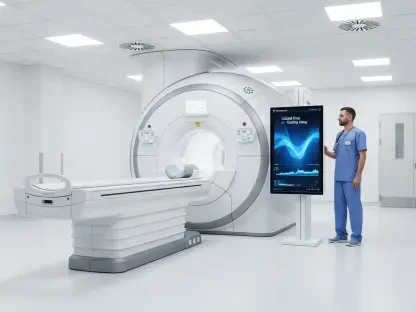

The field of pediatric surgery is currently witnessing a paradigm shift, moving away from purely mechanical repairs toward the biological reconstruction of complex organs. For children born with long-gap esophageal atresia, the challenge has always been the physical impossibility of connecting two ends of a food pipe that simply do not meet. In a landmark collaboration between Great Ormond Street Hospital and University College London, researchers have successfully engineered the first lab-grown esophagus that restores full function in large-animal models. This breakthrough utilizes a combination of decellularized donor tissue and the recipient’s own cells to create a living, growing graft. By bridging the gap between advanced bioengineering and clinical necessity, this technology offers a future where life-altering surgeries are replaced by personalized, regenerative solutions that grow alongside the patient.
The following discussion explores the intricate process of creating these bioengineered grafts, beginning with the transformation of pig donor tissue into a neutral scaffold and the subsequent repopulation of that structure with patient-derived muscle cells. We delve into the critical role of specialized bioreactors in fostering tissue integration and compare this regenerative approach to traditional, highly invasive surgeries that often result in lifelong gastrointestinal and respiratory complications. The conversation also highlights the long-term success seen in animal trials, where grafts developed their own nervous systems and blood supplies, and concludes with the logistical and technical milestones required to bring this “off-the-shelf” scaffold technology into human clinical trials within the next five years.
When using a pig donor esophagus as a base, how do you ensure the decellularization process completely removes animal cells while preserving the structural integrity? What specific mechanical properties must the scaffold maintain to support the eventual growth of new muscle and tissue?
The decellularization process is a delicate balancing act where we must act as molecular sculptors, stripping away every trace of the donor pig’s cellular identity while leaving the intricate “architecture” of the organ untouched. We use a series of chemical and physical washes to rinse out the porcine cells, ensuring that the remaining scaffold is a ghost-like, translucent tube of collagen and elastin that the recipient’s immune system will not recognize as foreign. It is vital that we do not over-process the tissue; if the chemicals are too harsh, we risk destroying the basement membrane, which is the essential “soil” where new cells must take root. The scaffold must retain its native elasticity and tensile strength because the esophagus is not just a passive tube; it is a dynamic organ that must withstand the constant internal pressure of swallowing. In our successful trials with eight large animals, we observed that maintaining this structural integrity allowed the graft to integrate fully within just three months, providing a stable foundation for the recipient’s own cells to organize into functional layers. By the six-month mark, the scaffold had transitioned from a synthetic-looking graft into a living tissue that was indistinguishable from the surrounding native esophagus in terms of both its look and its mechanical performance.
Once muscle cells are harvested from a patient biopsy, what steps are taken to multiply them in the lab? How does the one-week period inside a specialized bioreactor facilitate the integration of these cells into the scaffold before the graft is ready for implantation?
After we obtain a small biopsy of muscle tissue from the patient—often during a procedure they are already undergoing, such as the placement of a feeding tube—we isolate the muscle progenitor cells and begin a rigorous multiplication process in a controlled laboratory environment. These cells are nurtured in nutrient-rich media until they reach the high density required to “seed” the scaffold, a process that takes place over several weeks. Once we have a sufficient population, we inject these cells directly into the decellularized pig scaffold, but the real magic happens during the seven days the graft spends inside the bioreactor. This specialized chamber acts as an artificial womb, pumping vital growth fluids and nutrients through the tissue to simulate the natural environment of the body. This one-week period of “dynamic conditioning” is crucial because it encourages the cells to settle evenly across the scaffold, migrate into its pores, and begin the early stages of forming a cohesive muscle layer. The entire timeline from biopsy to a ready-to-implant graft is approximately two months, which perfectly aligns with the current clinical window where surgeons are preparing a child with long-gap esophageal atresia for their definitive repair.
Traditional surgeries for long-gap esophageal atresia often involve repositioning the stomach or intestines to bridge the gap. What are the primary long-term complications associated with these invasive procedures, and how does a lab-grown graft specifically address risks like breathing difficulties or gastrointestinal issues?
Traditional surgeries are often a case of “borrowing from Peter to pay Paul,” as they involve major anatomical rearrangements like the gastric pull-up, where the stomach is moved into the chest cavity to reach the upper esophagus. While these operations are life-saving, they come with a heavy toll; for instance, some children are born with gaps as large as 11 centimeters, making it nearly impossible to bridge the distance without extreme tension. This repositioning can lead to severe gastrointestinal reflux, a permanent risk of respiratory issues because the displaced organs press against the lungs, and even a long-term, unknown risk of cancer in the transposed tissue. We see the human cost in patients like Casey, a two-year-old who has spent half his life in the hospital and suffered vocal cord damage due to the sheer number of repeated, invasive surgeries. A lab-grown graft completely changes this equation by creating a “like-for-like” replacement that stays in the correct anatomical position, meaning the stomach remains in the abdomen where it belongs. By using an engineered esophagus, we avoid the trauma of moving other organs, which significantly reduces the likelihood of the breathing struggles and noise-making delays that many of these children currently face during their development.
In successful large-animal trials, engineered grafts eventually developed functioning nerves and blood vessels. How does using a recipient’s own cells eliminate the need for lifelong immunosuppression, and what metrics are used to confirm that the new tissue can contract with enough pressure for normal swallowing?
The most profound advantage of this technology is that the final graft is essentially “humanized” or “patient-specific” because it is repopulated with the recipient’s own muscle progenitor cells. Because the body recognizes these cells as “self” rather than “foreign,” we can completely bypass the need for lifelong immunosuppression, which is a massive relief for a growing child whose immune system needs to be robust to fight off everyday childhood illnesses. To verify that these grafts were truly functional, we utilized a cutting-edge technique called spatial transcriptomics to map the gene expression within the tissue layers; this confirmed that the genes being “turned on” were exactly those needed for a healthy, functioning esophagus. We also monitored the eight animals in our study to see if they could eat and grow at a healthy rate, which they did, proving that the graft had developed the necessary blood vessels and nerves to survive and communicate with the rest of the body. Most importantly, we measured the movement and pressure generated by the new esophagus, confirming it could produce contractions strong enough to squeeze food down toward the stomach with the same coordination as a natural organ.
If this technology transitions to human clinical trials within the next five years, how would hospitals manage a “bank” of donor scaffolds for different-sized children? What are the remaining technical hurdles to standardizing this manufacturing process for widespread pediatric use?
The vision for the next five years is to move from a bespoke research process to a standardized clinical pipeline where hospitals can access an “off-the-shelf” bank of decellularized pig scaffolds in various lengths and diameters. These scaffolds would be pre-cleansed and sterilized, ready to be personalized the moment a child is born with an esophageal defect, much like how pig heart valves have been used safely in cardiac surgery for over 50 years. However, several technical hurdles remain, such as the need to further automate the cell-seeding process and reduce the manual labor involved to ensure every graft is produced with the same high level of precision. We are also working on refining the technology to generate even longer grafts for the most severe cases and optimizing the initial blood flow to the graft immediately after surgery to ensure the tissue stays healthy in those first critical days. Standardizing this manufacture is not just about the biology; it is about creating a reliable, repeatable “blueprint” that can be scaled up so that a specialized center like Great Ormond Street Hospital can offer this as a routine alternative to the 180 babies born with esophageal atresia in the UK each year.
What is your forecast for the future of regenerative medicine in pediatric surgery?
I believe we are standing at the threshold of a “biological century” in pediatric surgery, where we will stop viewing the body as a collection of parts to be rearranged and start seeing it as a system capable of self-repair with the right bioengineered prompts. Within the next decade, the success we have seen with the esophagus will likely serve as a master key to unlocking similar treatments for other tubular organs, such as the trachea or the bowel, where children currently face equally devastating and invasive options. We will see a move toward “surgery-in-a-dish” protocols, where a child’s own cells are harvested at birth and grown into a replacement organ while the medical team manages their initial stability, leading to a single, definitive operation that lasts a lifetime. Ultimately, our goal is to eliminate the “patient for life” phenomenon, ensuring that children like Casey do not remember their time in the hospital as a series of miracles and setbacks, but as a single moment of healing that allowed them to grow, eat, and breathe without limits. This technology will turn the “impossible gap” into a bridge toward a normal childhood, proving that the most complex surgical problems are best solved by working with nature rather than trying to bypass it.