
For countless individuals who have undergone mastectomies, the journey toward physical and emotional recovery is often complicated by the limitations of existing reconstructive surgeries. A groundbreaking prototype of an injectable paste, developed by researchers from human skin cells and detailed in the journal ACS Applied Bio Materials, promises a paradigm shift in this field. This innovative biomaterial is engineered to serve as a space-filling agent following the surgical removal of cancerous tissue, presenting a less invasive, safer, and more cosmetically favorable option. It stands as a potential alternative to prevailing methods, such as prosthetic implants and autologous tissue transplantation, with the ultimate goal of improving long-term quality of life, comfort, and aesthetic satisfaction for patients navigating the difficult path of post-cancer care.
The Challenge of Post Mastectomy Reconstruction
Limitations of Current Surgical Options
The most common reconstructive techniques today present patients with a difficult choice between two complex surgical pathways, each with significant drawbacks. One major approach is autologous reconstruction, often called flap surgery, where skin, fat, and sometimes muscle are harvested from another part of the patient’s body—typically the abdomen, back, or thighs—to rebuild the breast mound. While this method can yield a result that looks and feels very natural, its primary disadvantage is the creation of a permanent and often substantial scar at the donor site. This is not merely a cosmetic concern; the procedure is essentially two major surgeries performed at once, increasing operative time, risk of complications like infection or tissue death, and leading to a longer and more painful recovery period. Patients must contend with healing at both the chest and the donor site, which can impact mobility and overall well-being for an extended time, making it an unsuitable option for some individuals.
In contrast, the other prevalent option involves the use of prosthetic implants, which circumvents the need for a secondary surgical site. However, this approach introduces a different set of risks associated with implanting a foreign object into the body. Beyond the immediate surgical risks of infection or hematomas, a significant long-term complication is capsular contracture. This occurs when the scar tissue, or capsule, that naturally forms around any implant tightens and hardens, squeezing the implant. This can cause chronic pain, a firm or unnaturally round appearance, and distortion of the breast’s shape. Correcting capsular contracture often requires one or more additional surgeries to release the scar tissue or replace the implant entirely. For many patients, the prospect of ongoing surgical interventions and the potential for an unnatural aesthetic outcome make implants a less-than-ideal solution, highlighting the urgent clinical need for a less burdensome alternative.
A Novel Biomaterial Solution
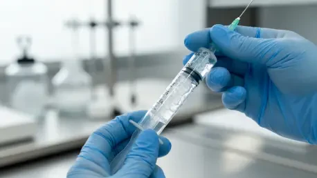
Addressing these significant clinical challenges, a research team led by Pham Ngoc Chien and Chan-Yeong Heo has pioneered an injectable form of an acellular dermal matrix (ADM). This sophisticated biomaterial is derived from human skin that has undergone a critical processing step called decellularization, where all original cells are carefully removed. This process is essential as it eliminates the risk of an immune rejection by the recipient’s body, which would otherwise identify the material as foreign and attack it. What remains is the skin’s essential structural scaffold, a rich network of proteins like collagen and elastin, along with various growth factors that are vital for healing and tissue regeneration. While ADM is already a valuable tool in medicine, it is typically available in a solid sheet form, used for applications like tendon repair or as a supportive sling in certain plastic surgery procedures, requiring surgical implantation and shaping.
The key innovation presented by the research team lies in the transformation of this established biomaterial into a thick, injectable paste specifically designed to fill the irregular voids left by tumor removal in a minimally invasive manner. The meticulous process began with a skin sample donated by a living female participant. This tissue was first decellularized before being frozen and pulverized into fine ADM particles. These particles were then carefully rehydrated with water to achieve a paste-like consistency suitable for injection through a syringe. To validate the safety and efficacy of this novel formulation, the team conducted a comprehensive preclinical study. They injected small amounts of the ADM paste into rats, and over a six-month observation period, critically compared its performance against two commercially available ADM products that are currently used in clinical settings, setting a high bar for their prototype.
Promising Results and Future Potential
Preclinical Findings Demonstrate Superiority
The preclinical study conducted over a six-month period yielded exceptionally promising findings, with the primary outcome being the excellent safety profile of the newly developed ADM paste. Throughout the entire study, the rats that received injections of the material exhibited no adverse health effects, a clear indication that the paste is highly biocompatible and well-tolerated by a living biological system. This is the first and most critical hurdle for any new implantable medical device or material, as a negative host response could lead to chronic inflammation, tissue damage, or systemic illness. The absence of such reactions suggests that the decellularization and preparation process was successful in creating a material that the body accepts rather than rejects, paving the way for further investigation into its regenerative capabilities and long-term performance as a soft tissue filler in a clinical setting.
A crucial and superior outcome was observed in the body’s reaction to the implanted paste compared to existing commercial products. The tissue layers, known as fibrous capsules, that naturally formed around the researchers’ injectable material were significantly thinner than those that developed around the commercially available ADMs. In reconstructive surgery, a thinner, more flexible capsule is highly desirable, as it signifies a milder inflammatory response and better integration of the biomaterial with the host tissue. Thick, dense capsules are a clinical red flag, strongly associated with an increased risk of long-term complications, most notably capsular contracture. This condition can cause pain, hardening, and a visible distortion of the breast mound, often necessitating corrective surgery. The thinner capsules observed in this study suggest a substantially lower risk of such complications, pointing toward a safer and more stable long-term result for patients.
The Path from Laboratory to Clinic
The researchers articulated a multifaceted vision for this injectable matrix, framing it not as a passive filler but as an active regenerative scaffold. Its design was intended to promote the growth of new blood vessels, a process known as angiogenesis, which is essential for the material’s survival, integration, and eventual replacement by the patient’s own tissue. Furthermore, it was engineered to encourage positive tissue remodeling while actively minimizing inflammation and significantly reducing the risk of capsular contracture. The collective impact of these properties, as envisioned by the team, could make breast reconstruction safer, less invasive, and more accessible. However, the researchers acknowledged that this work represented a crucial but foundational first step. Before the injectable ADM paste could be considered for clinical use in humans, it had to undergo further extensive evaluation, including longer-term safety trials and more complex tests in larger animal models that more closely mimic human anatomy. These future studies were deemed essential to confirm its long-term efficacy and predictable safety profile before its potential transition from the laboratory to the operating room.


