
The primary obstacle to curing many aggressive forms of cancer is not a lack of potent chemical agents but rather the physical inability to smuggle these treatments past the cellular gatekeepers of the body. Most revolutionary therapies, designed to dismantle the very foundation of a tumor, are composed of large, complex molecules that are naturally repelled by the oily, protective lipid bilayer of a cell membrane. This biological standoff often forces clinicians to use higher, more toxic doses of medication in the hope that a small fraction will accidentally seep into the target cells. However, a pioneering development from researchers at Duke University, known as Sonoporation-assisted Precise Intracellular Nanodelivery (SonoPIN), is fundamentally altering this dynamic by using acoustic energy to mechanically bypass cellular defenses. By synchronizing the physical behavior of microbubbles with high-frequency sound waves, this technology creates a temporary, non-invasive portal that allows life-saving drugs to enter exactly where they are needed most.
Overcoming Biological Barriers Through Acoustic Force
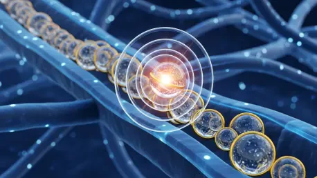
The Mechanism: Controlled Sonoporation
The operational core of the SonoPIN system relies on a phenomenon called sonoporation, which transforms standard medical microbubbles into active delivery tools. These gas-filled spheres, which have been used safely in diagnostic imaging for years, respond dynamically when they are struck by specifically tuned ultrasound waves. Rather than simply reflecting the sound to create a picture, the microbubbles undergo a series of rapid, violent oscillations that culminate in a localized collapse. This physical event is not a chaotic explosion but a calculated release of energy that generates microscopic jets of fluid and intense shock waves. These forces exert just enough pressure on the nearest cell membrane to create nanoscopic, temporary punctures. This mechanical breach effectively turns the cell’s outer wall into a permeable sieve for a brief window of time, allowing large therapeutic molecules to flood into the cytoplasm before the membrane naturally seals itself.
The brilliance of this approach lies in its ability to facilitate the entry of “bulky” drugs like PROTACs, which are typically too large to cross the membrane through traditional diffusion. Because the lipid bilayer is inherently fluid and self-healing, the cells can survive this momentary intrusion without suffering permanent structural damage. This represents a significant departure from chemical delivery methods that often rely on toxic solvents or viral vectors to gain entry. By using purely mechanical force, the researchers have created a delivery “key” that is independent of the drug’s chemical composition. Consequently, the SonoPIN platform can be adapted to transport a wide variety of therapeutic cargoes, ranging from traditional chemotherapeutics to advanced protein degraders, all while ensuring that the cellular machinery remains intact and functional once the drug has been successfully delivered and the membrane has closed.
Precision Engineering: Molecular Targeting and Safety
While the mechanical power of ultrasound provides the means of entry, the true innovation of SonoPIN is its ability to distinguish between a malignant tumor and a healthy organ. To achieve this, the Duke team decorated the surface of the microbubbles with synthetic nucleic acid strands that function as a biological GPS. These strands are custom-engineered to recognize and bind only to specific protein receptors that are overexpressed on the surface of cancer cells. When these modified microbubbles are injected into the bloodstream, they ignore healthy tissues and naturally cluster around the cancerous mass. This localized accumulation ensures that when the ultrasound wand is activated, the resulting sonoporation only occurs at the site of the disease. This spatial control is crucial for protecting the patient from the systemic side effects that often make modern cancer treatments as debilitating as the illness itself.
The safety profile of this system is further enhanced by the precision of the ultrasound trigger, which allows doctors to “turn on” the drug delivery only within a clearly defined three-dimensional space. Even if some microbubbles remain in the general circulation, they stay inert and harmless unless they are directly hit by the focused acoustic beam. This dual-layer of protection—biological targeting combined with physical activation—effectively shields the rest of the body from high-potency drugs that would otherwise cause widespread damage. The result is a treatment environment where 99% of non-targeted healthy cells remain completely viable and unaffected, even when they are in close proximity to the treated area. This high degree of selectivity enables the use of much more aggressive therapeutic agents, as the risk of off-target toxicity is virtually eliminated through the localized nature of the acoustic activation.
Validating Efficacy and Future Clinical Potential
Experimental Results: Efficacy in Laboratory Testing
The data gathered during benchtop testing provides a compelling case for the immediate adoption of SonoPIN in advanced oncological research. In controlled experiments published in the Proceedings of the National Academy of Sciences, the researchers observed that cells treated with the SonoPIN method exhibited a seven-fold increase in drug uptake compared to those subjected to conventional delivery protocols. This massive surge in intracellular concentration led to a rapid therapeutic response, with 50% of the targeted cancer cells undergoing programmed cell death, or apoptosis, within just sixty seconds of ultrasound exposure. These results are particularly impressive given that the drugs used, such as BRD4-targeting PROTACs, are notoriously difficult to deliver effectively. The ability to achieve such high mortality rates in a tumor population in under a minute highlights the sheer efficiency of using mechanical force over biochemical persuasion.
Beyond the raw numbers of cell death, the researchers utilized fluorescent markers to track the movement of the drug molecules in real-time. This visualization confirmed that the PROTACs were not just sticking to the outside of the cells but were being successfully driven deep into the interior, where they could immediately begin dismantling the proteins necessary for cancer survival. The experiments also confirmed the “Goldilocks zone” of ultrasound intensity—a specific frequency that maximizes membrane permeability without triggering irreversible cell lysis. This delicate balance ensures that the treatment is lethal to the cancer’s internal structures while remaining gentle on the cell’s overall integrity during the delivery process. Such refined control suggests that the platform is ready for more complex biological environments, providing a reliable foundation for the next stage of development in living organisms.
Future Horizons: Mechanical Pharmacology and Beyond
The successful validation of SonoPIN opens the door to a new era of “mechanical pharmacology,” where the physical properties of sound and matter are used to solve the most stubborn problems in medicine. Because the delivery mechanism is purely physical, it is not limited by the specific biochemistry of the drug being transported. This suggests that the platform could eventually be used to deliver gene-editing complexes like CRISPR/Cas9, which are even larger and more fragile than PROTACs. In a clinical setting, this would allow for the precise editing of a patient’s DNA within a specific organ without the risks associated with systemic viral delivery. The versatility of the microbubble platform means that by simply swapping the synthetic targeting strands, the same technology could be repurposed to treat a vast array of conditions beyond oncology, including genetic disorders and localized infections.
As the research transitions from laboratory benches to animal models, the focus will shift toward optimizing the intravenous delivery and the precision of the ultrasound wands used in a hospital environment. The long-term vision involves a streamlined procedure where a patient receives a quick injection of targeted microbubbles and therapeutic agents, followed by a brief, painless ultrasound session. This approach would allow for maximum drug concentration at the tumor site while maintaining near-zero levels in the rest of the circulatory system. To realize this potential, future efforts should prioritize the standardization of microbubble manufacturing and the development of portable, high-precision ultrasound arrays. By integrating these physical tools with the latest breakthroughs in synthetic biology, the medical community can finally move toward a future where “undruggable” targets become a relic of the past, replaced by localized, high-potency cures.


