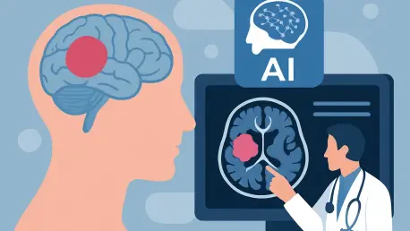

What if a surgeon could instantly determine, mid-operation, whether a brain tumor is a deadly glioblastoma or a treatable lymphoma that demands an entirely different approach? This question isn’t just a thought experiment—it’s a life-or-death reality for thousands of patients undergoing brain surgery each year, where diagnostic errors can drastically alter the course of treatment and survival. With the integration of artificial intelligence (AI) into the operating room, a groundbreaking possibility emerges. A tool named PICTURE is leading this charge, promising to redefine precision in neuro-oncology by providing real-time insights when seconds count.
Why Brain Tumor Diagnosis Is a High-Stakes Challenge
The operating room is a place of intense pressure, where every decision can tip the balance between life and loss. Brain tumors like glioblastoma, an aggressive cancer, and primary central nervous system lymphoma (PCNSL), a rarer immune-cell cancer, often appear strikingly similar under quick microscopic review. Yet, their treatments diverge sharply—surgical removal for one, chemotherapy for the other. Misdiagnosis, occurring in about 5% of rapid assessments, can lead to devastating outcomes, such as unnecessary operations or delayed critical care. This diagnostic dilemma, compounded by the time constraints of surgery, underscores a pressing need for faster, more reliable tools.
The stakes extend beyond individual cases to a systemic level. Thousands of patients globally face these uncertainties annually, with traditional frozen-section analysis—used during surgery—often distorting cellular details and taking up precious minutes. Final pathology reports, available days later, can contradict initial findings, leaving surgeons to second-guess decisions made under duress. Addressing this gap isn’t just about improving outcomes; it’s about transforming trust in surgical precision at a moment when there’s no room for error.
Unveiling PICTURE: AI’s Answer to Diagnostic Uncertainty
At the forefront of this medical revolution stands PICTURE, or Pathology Image Characterization Tool with Uncertainty-aware Rapid Evaluations, developed by researchers at Harvard Medical School. Tested on over 2,000 brain pathology slides across five international hospitals, this AI tool has demonstrated a remarkable 98% accuracy in distinguishing glioblastoma from PCNSL during rapid assessments. Unlike existing methods, it delivers results in real time, aligning with the urgent pace of brain surgery and empowering surgeons to make informed choices on whether to operate or pivot to alternative treatments.
What makes this technology stand out is not just its precision but its adaptability. Beyond the two primary tumors, PICTURE can identify 67 other central nervous system cancers, positioning it as a versatile diagnostic companion. Its unique uncertainty detector further enhances reliability by flagging ambiguous cases for human review, acknowledging the complexity of over 100 brain tumor subtypes. This balance of automation and human oversight marks a significant leap toward safer, more confident decision-making in high-stakes environments.
Real-World Impact: Stories and Statistics That Speak Volumes
Voices from the medical field amplify the transformative potential of this innovation. Senior author Kun-Hsing Yu from Harvard Medical School emphasizes, “This tool isn’t designed to replace pathologists but to equip them with critical data during the most pivotal moments of surgery.” Published findings in a leading scientific journal reveal that in complex cases—where human experts disagree, leading to misdiagnosis rates as high as 38%—PICTURE consistently offers clarity, often surpassing seasoned professionals in accuracy.
Testimonies from testing sites paint a vivid picture of its impact. In a resource-limited hospital, a surgeon used the tool to confirm a tumor type that would have otherwise taken days to diagnose, potentially sparing a patient from an invasive and unneeded procedure. Supported by the National Institutes of Health, the tool’s validation across diverse international datasets reinforces its credibility. These real-world applications highlight not just technological prowess but a tangible difference in patient care, bridging gaps where time and expertise are scarce.
Bridging Gaps: Practical Uses in Today’s Operating Rooms
Integrating AI like PICTURE into surgical practice offers immediate and far-reaching benefits. In the heat of an operation, it provides surgeons with instant tumor identification, ensuring that treatment—whether aggressive resection for glioblastoma or non-surgical options for PCNSL—matches the cancer’s true nature. This capability can drastically reduce errors, aligning decisions with the best possible outcomes during critical windows of opportunity.
Beyond individual surgeries, the tool holds promise for leveling disparities in healthcare access. In regions lacking neuropathology specialists, it can serve as a virtual expert, delivering high-level diagnostics where human resources are limited. Additionally, its potential as a training platform for aspiring pathologists is immense, offering guided analysis of subtle brain lesion differences. These applications suggest a future where technology not only enhances precision but also democratizes specialized care on a global scale.
Looking Ahead: Expanding Horizons and Overcoming Limits
The journey doesn’t end with current achievements; there’s a clear path for growth. Expanding research to validate PICTURE across diverse patient demographics remains essential, especially since initial testing involved a predominantly white sample. Broadening its scope to include additional tumor types and integrating molecular data could further refine its accuracy, addressing nuances that visual analysis alone might miss. These steps are crucial for ensuring equitable and comprehensive impact.
Collaboration between technology developers and medical communities has proven vital in shaping this tool’s success. Encouraging further studies from 2025 onward will solidify its role in neuro-oncology, potentially setting a standard for AI-assisted diagnostics. Reflecting on past efforts, the dedication to balancing innovation with ethical safeguards, like uncertainty detection, stands as a cornerstone. Moving forward, stakeholders must prioritize accessibility and continuous improvement, ensuring that such advancements reach every corner of healthcare, transforming brain surgery into a field defined by precision and hope.


