3D bioprinting is an innovative technology that promises to revolutionize cancer research and treatment by providing more accurate and complex models of tumor tissue. This advancement is particularly significant in the study of cancers involving the BRCA1 gene. Tumor modeling with 3D bioprinting mimics the complex architecture and microenvironment of cancer tissue, paving the way for improved understanding of tumor biology, more effective drug testing, and the development of personalized cancer treatments. This transformative approach aims to address the limitations of traditional cancer models, which have long been criticized for their oversimplification and lack of reproducibility.
The Limitations of Traditional Cancer Models
Flat Cultures and Organoids: A Simplistic Approach
Traditional cancer models, such as flat cultures, fall short in representing the multifaceted interactions that occur within the tumor microenvironment. These flat cultures offer a two-dimensional perspective, lacking the depth needed to capture the interplay between different cell types, such as immune cells, blood vessels, and the extracellular matrix. This deficit hinders researchers’ ability to study the full scope of tumor growth and progression. While organoids provide a more three-dimensional approach, they too come with limitations. Organoids can replicate the architecture of the tissue to some degree but often lack important components and can be inconsistent in their formation.
The tumor microenvironment is a dynamic and intricate network. The lack of complexity in traditional models limits their effectiveness in gaining true insights into how tumors develop, interact with surrounding tissues, and respond to treatments. Without a comprehensive model, the research risks missing critical aspects of these processes, leading to incomplete or inaccurate findings. As a result, there has been a growing demand for more sophisticated cancer models that can faithfully represent the conditions within a human body, leading researchers to explore advanced techniques like 3D bioprinting to bridge this gap.
The Need for a More Complex Model
Given the intricate nature of the tumor microenvironment, a more complex and comprehensive model is necessary for accurate cancer research. Traditional flat cultures and even organoids do not sufficiently encapsulate the variability and interaction of various cell types and structures, making it challenging to obtain reliable and applicable data. This oversight can result in misleading outcomes, particularly in understanding how tumors evolve and respond to therapies.
The necessity for sophisticated models becomes even more evident when considering the dynamic processes within tumors. Tumors are not static entities; they evolve by interacting with their surroundings, recruiting different cells, and undergoing genetic mutations over time. Traditional models fail to replicate these dynamic processes, resulting in underestimations of tumor resilience and drug resistance. There is an urgent need for advanced modeling techniques like 3D bioprinting, which can create more accurate representations of the human tumor microenvironment, providing a platform for better research and more effective drug development.
The Promise of 3D Bioprinting
Building Tissue Structures Layer by Layer
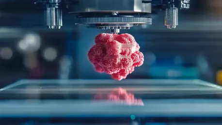
3D bioprinting has the potential to revolutionize cancer research by constructing tissue structures layer by layer using bioinks composed of living cells, active molecules, or biomaterials like alginate or collagen. This innovative approach allows for the creation of tissue models that closely mimic the natural mechanical and biological properties of the target tissue. By using bioinks, researchers can replicate the various cell types and the extracellular matrix found within the tumor microenvironment, producing a more accurate and complex model for study.
The precision of 3D bioprinting offers superior control over the spatial arrangement of different cell types within the printed tissue. This ability to apportion cells in designated areas enhances the realism of the model, allowing for detailed examination of cell-cell interactions, tumor growth, and drug responses. Researchers can extend this precision to include components like blood vessels, essential for studying tumor vascularization and metastasis. This high fidelity in modeling provides a richer and more accurate platform for studying cancer, thereby promising more effective and efficient research.
Precision and Control in Tumor Modeling
One of the most significant advantages of 3D bioprinting is the unparalleled control it offers researchers in modeling tumors. Unlike traditional 3D models, 3D bioprinting allows for precise spatial arrangement of various cell types, enabling a more realistic mimicry of tumor architecture. This detailed control is vital for studying intricate cell-cell interactions within the tumor microenvironment, yielding more accurate insights into the behavior of tumors, their growth patterns, and their response to treatments.
Furthermore, bioprinted models can incorporate essential components like blood vessels, offering a deeper understanding of how tumors stimulate vascularization—a critical process for tumor growth and metastasis. The inclusion of these elements makes bioprinted models a superior tool for cancer research, as they provide a more holistic view of tumor biology. This advanced level of detail and realism not only aids in developing new cancer therapies but also in refining existing treatments by providing a more accurate testing ground.
Case Studies and Research Highlights
Stephanie Willerth’s Work on Neural Tissue Models
Stephanie Willerth, a prominent biomedical engineer at the University of Victoria, has significantly contributed to the field of tissue engineering using 3D bioprinting. Her team’s research has primarily focused on creating 3D neural tissue models for studying neurological disorders. However, they have increasingly turned to bioprinting technologies to enhance the precision and reproducibility of their tissue models. By meticulously placing specific cell types in designated areas, Willerth’s team has managed to construct more accurate and consistent models, representing a substantial advancement in neurological research.
Willerth’s innovative approach allows for a higher degree of control over the placement and interaction of cells, leading to more detailed and reliable models. This precision is particularly valuable for studying the complex interplay of cells within the tumor microenvironment. In their latest efforts, her team has utilized these techniques to build models of glioblastoma—a highly aggressive form of brain cancer. Their work demonstrates the potential of bioprinting to improve the study of cancer cell behavior and response to treatments, providing a more robust platform for developing targeted therapies.
İbrahim Özbolat’s Breast Tumor Model
İbrahim Özbolat, a researcher at Pennsylvania State University, has developed an intricate bioprinted breast tumor model to study cancer more effectively. His model incorporates various cell types and vasculature, mimicking the complexity found within actual tumors. This advanced model is particularly used to explore how CAR T cell therapies, effective against blood cancers, can be optimized for treating solid tumors. By providing a detailed replica of the tumor microenvironment, Özbolat’s work highlights the challenges CAR T cells face in navigating and infiltrating dense tumor structures.
The insights gained from Özbolat’s bioprinted models are invaluable for enhancing the effectiveness of CAR T cell therapies. His research underscores the need to understand the physical barriers and interactions within the tumor microenvironment that can impact treatment efficacy. Through his innovative use of 3D bioprinting, Özbolat is paving the way for the development of more effective cancer immunotherapies, offering hope for improved outcomes in patients with solid tumors. His work exemplifies the transformative potential of bioprinting in advancing cancer research.
Challenges and Limitations
Temporal Interactions and Tumor Evolution
Despite the significant advantages of bioprinted models, they are not without their challenges. One major limitation is the inability to replicate the temporal interactions that occur during the gradual growth of a tumor. Tumors in the body evolve over time, recruiting various cell types from their surroundings, and this dynamic process is difficult to capture with current bioprinting technology. While bioprinted models can effectively reproduce the spatial arrangement of cells, they fall short in mimicking the evolving nature of tumor biology.
The inability to match the temporal dynamics of tumor development can lead to gaps in understanding how tumors respond to different stages of growth and treatment. This limitation highlights the need for further advancement in bioprinting technologies. Future innovations must address this challenge by integrating mechanisms that enable the continuous evolution of the bioprinted models, thereby providing a more accurate representation of tumor progression and interaction with treatments over time. Continued research and development in this area are crucial for overcoming these limitations and enhancing the utility of bioprinted models in cancer research.
Ethical and Practical Considerations
The use of animal models has long posed significant ethical and practical challenges in cancer research. Ethical considerations aside, animal models often fail to accurately represent human physiology, leading to potential discrepancies in research findings and drug development. 3D bioprinted models offer a promising alternative by providing a more human-relevant platform for experimentation. These models incorporate healthy human tissue, reducing the reliance on animal testing, and offering a more ethically sound approach to research.
Bioprinted models also present practical benefits by enabling greater control over experimental conditions and reducing overall experimentation time and costs. The ability to tailor the models to specific research needs and conditions enhances reproducibility and reliability in results. While bioprinted models are not perfect, they represent a significant step forward in creating more ethical, efficient, and accurate tools for cancer research. Addressing the remaining limitations through continued innovation will further solidify their potential to replace animal models in various aspects of cancer research and drug development.
Implications for Cancer Drug Development
High-Throughput Drug Screening
The success rate of clinical trials in cancer drug development has historically been low, with many promising drugs failing to demonstrate efficacy in human trials. 3D bioprinted tumor models hold significant promise in improving this success rate by offering a more accurate platform for high-throughput drug screening. These models closely mimic the complexities of human tumors, providing a more representative environment for testing the efficacy and safety of potential treatments.
Drugs like cisplatin and 5-fluorouracil, which have shown effectiveness in 2D cultures, often face reduced efficacy in more complex 3D models and animal tests. Bioprinted models can bridge this gap by offering a more realistic testing ground, reconciling discrepancies between preclinical and clinical results. The use of these advanced models can streamline the drug development process, enabling faster transitions to animal testing and clinical trials with greater confidence in the treatments’ potential success. This advancement not only accelerates the development of new cancer therapies but also enhances the likelihood of successful outcomes in clinical applications.
Personalized Cancer Treatments
3D bioprinting is a groundbreaking technology that holds the potential to revolutionize cancer research and treatment by offering more precise and intricate models of tumor tissue. This technological leap forward is particularly impactful for studying cancers associated with the BRCA1 gene. By employing 3D bioprinting, researchers can replicate the complex architecture and microenvironment of cancer tissue, which significantly enhances our understanding of tumor biology. This innovative method facilitates more effective drug testing and paves the way for developing personalized cancer treatments.
The traditional models of cancer have long been criticized for their overly simplistic nature and lack of reproducibility. These conventional methods often fail to capture the intricate dynamics and heterogeneity of tumor tissues found in the human body. In contrast, 3D bioprinting offers a more realistic and detailed representation of the tumor environment, addressing these critical limitations.
Ultimately, this transformative approach promises to advance therapeutic strategies and cancer treatments by enabling more precise modeling of tumors, leading to better understanding, more reliable drug efficacy testing, and tailored treatment options for patients. With these advancements, the future of cancer research and treatment could be profoundly more effective and personalized, offering new hope to patients and healthcare professionals alike.


