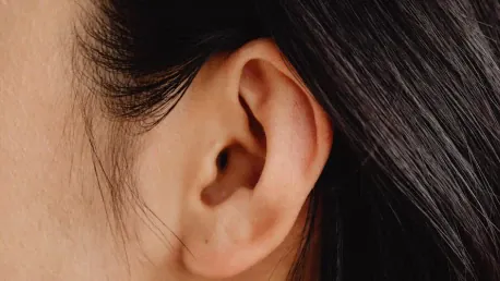
Ear reconstruction is one of the most challenging tasks in reconstructive surgery, demanding both structural integrity and aesthetic appeal. Recreating the outer ear, or auricle, involves achieving both structural integrity and aesthetic appeal, a complex endeavor even for an experienced surgeon. Traditional methods have primarily relied on synthetic materials or rib cartilage, which come with numerous inherent challenges. Recent advancements in 3D printing and tissue engineering, however, are proving to be game-changers in this field, offering new possibilities for more effective and natural-looking ear reconstructions.
The Traditional Challenges in Ear Reconstruction
Ear reconstruction typically involves the laborious process of using synthetic materials or harvesting cartilage from a patient’s ribs. This method requires surgeons to possess a high level of anatomical expertise and artistic skill to sculpt these materials into the precise shape of an ear. Despite these efforts, achieving the necessary structural fidelity and natural appearance remains a significant challenge. Limitations such as the rigidity of the materials and the lack of integration with the body’s natural tissues often lead to suboptimal outcomes, both aesthetically and functionally.
Surgeons must meticulously carve rib cartilage to replicate the intricate shapes required for an auricle, which is not only time-consuming but also highly dependent on the individual surgeon’s skill. Moreover, utilizing synthetic materials can result in complications like infections or rejection by the body, making the procedure less reliable. Even in the best hands, traditional methods might fail to deliver the desired level of naturalness and functionality, leaving patients with less satisfactory results.
Enter 3D Printing and Tissue Engineering
In a bid to overcome these significant limitations, Jason Spector, a plastic and reconstructive surgeon at Weill Cornell Medicine, has pioneered a revolutionary approach to ear reconstruction. Leveraging his extensive expertise in tissue engineering and the innovative power of 3D printing, Spector and his team aim to redefine the standards of this challenging surgical procedure. By utilizing 3D printing technologies, they have successfully created highly accurate models of human ears using polylactic acid bioink, which serves as a scaffolding material.
These 3D-printed models provide a structural template designed to support tissue growth. According to a groundbreaking proof-of-concept study published in Acta Biomaterialia, these scaffolds can effectively support the development of new cartilage tissue once implanted into rodents. This blend of structural precision offered by 3D printing and the organic growth fostered by biological materials represents a balanced and effective strategy for creating more natural-looking and functional ear reconstructions.
The Proof-of-Concept Study
Spector’s team has demonstrated the viability and efficiency of combining 3D printing technology with decellularized cartilage pieces. These constructs were implanted into rodents, where they successfully fostered the development of new cartilage tissue. This innovative approach allowed the constructs to benefit from both the precise structural accuracy provided by 3D printing and from the biological signals necessary for effective tissue formation, derived from the decellularized cartilage pieces.
Experts in the field have praised the study for its innovative combination of synthetic and biological materials. Jason Burdick from the University of Colorado Boulder commended the effective amalgamation of 3D-printed scaffolds and biological tissues, underscoring the potential this method holds in harnessing tissue regeneration more efficiently. Additionally, Stephanie Willerth from the University of Victoria hailed the six-month persistence and cellular infiltration of the constructs as significant milestones, further validating the approach and its promise for future applications in human ear reconstruction.
Addressing Scalability and Cost Challenges
Despite its promising results, this novel method still faces several significant hurdles, including the scalability of printing human-sized organs and the financial costs involved. Scaling up from rodent-sized constructs to human-sized ears is a complex process that demands significant resources and technological advancements. The transition from small-scale experimental models to full-scale clinical applications involves numerous scientific and logistical challenges that must be addressed before this technology can be widely adopted.
Moreover, the financial costs of culturing human cells on a large scale also pose a considerable challenge. The bioinks and tissues used in these constructs are currently expensive, which makes widespread clinical application financially impractical at the moment. However, continuous advancements in 3D printing technologies and tissue engineering could eventually reduce these costs, making such procedures more accessible and affordable. It is therefore crucial to push forward with research and development to overcome these obstacles and pave the way for broader adoption of these innovative techniques.
Personalized Medical Treatment and Future Directions
One of the most revolutionary aspects of using 3D-printed ear scaffolds is the potential for highly personalized medical treatment. By employing 3D scans of a patient’s existing ear structures, surgeons can create custom-fit models that precisely match the individual’s anatomical requirements. This level of personalization promises to deliver better aesthetic outcomes and improved functional results, significantly enhancing the overall quality of ear reconstructions.
Looking to the future, Spector’s plans include experimenting with different bioinks that can degrade more quickly after implantation, thereby allowing the body’s natural tissue to replace the scaffold more effectively. Another key area of focus is enhancing the scaffold materials to support the collagen elasticity necessary for a more natural tissue structure. By refining these bioinks and scaffold designs, researchers are working towards achieving reconstructions that are not only aesthetically pleasing but also functionally sound, closely mimicking the flexibility and resilience of natural human ears.
Conclusion
Ear reconstruction ranks among the most challenging endeavors in reconstructive surgery, requiring a blend of structural robustness and visual appeal. Crafting the outer ear, or auricle, is a demanding process, even for seasoned surgeons, due to the necessity of achieving both strength and aesthetics. Traditional techniques typically use synthetic materials or rib cartilage, but these methods present numerous challenges and limitations. In recent years, advancements in 3D printing and tissue engineering have emerged as revolutionary tools in this field. These cutting-edge technologies are opening new avenues for creating ear reconstructions that are not only more effective but also more natural-looking. This innovation holds promise for improving both the procedural success rate and the quality of the reconstructed ear, offering hope to patients requiring these complex surgeries. As these new methods evolve, they are setting new standards and creating better outcomes in the realm of ear reconstructive surgery, making what was once a highly challenging task more feasible and successful.


