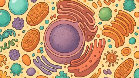

Imagine stepping into a bustling concert venue where every inch is packed with enthusiastic fans, movement is slow, and personal space is nearly nonexistent, yet somehow, everything functions with a strange kind of order. This vivid scene mirrors the surprising reality of the cytoplasm inside the cells of multicellular organisms like the tiny worm Caenorhabditis elegans. Far from the outdated notion of a fluid, pool-like environment where molecules swim freely, recent research reveals a cellular interior that is densely crowded and highly organized. A groundbreaking study from UC Davis, published in Science Advances, has shifted the understanding of cellular dynamics by examining the biophysical properties of cytoplasm in a living animal. This work highlights how molecular crowding and compartmentalization profoundly affect critical processes such as molecule movement, drug delivery, and disease progression, challenging long-held assumptions derived from single-celled models.
Unveiling a New Cellular Reality
The traditional view of cytoplasm as a watery, open space has been upended by findings that showcase its complexity within multicellular organisms. In C. elegans, researchers discovered that the cellular interior operates under intense spatial constraints, with particle movement slowed by as much as 50 times compared to single-celled systems like yeast. This stark difference arises from the crowded nature of the environment, where molecules are packed tightly, much like attendees at a sold-out show. The UC Davis team, including doctoral candidate Xiangyi Ding and professors Daniel Starr and G.W. Gant Luxton, emphasized that studying cells in a living organism provides a more accurate picture of biological behavior. Their work suggests that past research using isolated cell cultures fails to capture the true dynamics at play, urging a reevaluation of how cellular processes are understood and investigated in real-world contexts.
Beyond the sheer density, the study revealed that the cytoplasm in these worms is not just crowded but also highly compartmentalized. This means that molecules and particles are restricted to specific zones within the cell, unable to roam freely as previously thought. Such organization implies a level of control that is critical for cellular function, affecting everything from nutrient transport to stress responses. By focusing on a living model, the research underscores the limitations of simpler systems and highlights the necessity of in vivo studies to grasp the intricate balance of cellular life. This shift in perspective could have far-reaching implications for fields like pharmacology, where understanding molecule movement is key to effective drug design and delivery.
Tools and Techniques Behind the Discovery
To uncover these hidden aspects of cellular environments, the UC Davis team employed an innovative approach using Genetically Encoded Multimeric Nanoparticles (GEMs). These tiny tools, roughly the size of ribosomes at about 40 nanometers, were engineered with fluorescent tags and integrated into the genome of C. elegans. High-speed time-lapse microscopy allowed precise tracking of GEM movement within intestinal and skin cells, taking advantage of the worm’s transparent skin for clear observation. The results were striking, showing not only a dramatic slowdown in particle motion due to crowding but also a clear restriction to specific cytoplasmic regions. This method marked a significant departure from earlier studies on yeast or mammalian cell cultures, which showed no such spatial limitations, and opened new avenues for exploring cellular dynamics in a living context.
Further analysis using GEMs provided insights into the unexpected constraints within the cytoplasm, revealing a level of organization previously unseen in simpler models. The transparency of C. elegans proved invaluable, offering a window into real-time cellular activity that cell cultures cannot replicate. This technological advancement, though complex and time-intensive to develop, has proven its worth by delivering data that challenges existing paradigms. The ability to visualize and quantify how particles navigate such a dense and structured environment lays the groundwork for deeper investigations into how cells manage internal space. Such tools could redefine research methodologies, pushing scientists to prioritize living systems over isolated ones for a truer understanding of biological processes.
Mechanisms of Crowding and Control
Delving into the reasons behind this crowded and compartmentalized state, the research pinpointed two key mechanisms at work in C. elegans cells. Ribosomes, acting like dense packing material, contribute significantly to the crowded environment, slowing down particle movement through sheer concentration. Simultaneously, the protein ANC-1 functions as a structural scaffold, maintaining orderly compartmentalization by restricting particles to designated areas. Experiments disrupting ANC-1 production showed increased mobility of GEMs despite the density, while altering ribosome levels further enhanced movement. This dual system of control illustrates a sophisticated balance that cells in multicellular organisms maintain, a complexity not fully appreciated in studies of single-celled or cultured systems.
The interplay between ribosomes and ANC-1 offers a glimpse into how cells adapt to their packed interiors while ensuring functionality. This balance is crucial for processes like protein synthesis and cellular repair, which rely on precise spatial organization. The findings suggest that cellular environments in living organisms are far more regulated than previously thought, with each component playing a distinct role in managing space. Understanding these mechanisms could inform strategies for tackling diseases where cellular crowding or misorganization plays a role, such as neurodegenerative conditions. As research progresses, these insights may guide the development of therapies that target specific cellular constraints, enhancing treatment precision.
Future Horizons in Cellular Research
Looking ahead, the UC Davis team is eager to expand the application of GEMs to other cell types within C. elegans, such as neurons, to explore how cytoplasmic properties evolve during aging and neurodegeneration. Plans are also underway to adapt this methodology for more complex organisms like zebrafish, broadening the understanding of cellular dynamics across species. The potential to uncover variations in crowding and compartmentalization in different biological contexts is immense, offering clues about how cellular environments influence health and disease. Such studies could reveal whether the principles observed in worms hold true in higher organisms, paving the way for universal insights into cellular behavior.
The development of GEMs for multicellular research, while challenging, represents a significant leap forward in scientific capability. This tool’s ability to provide detailed, real-time data on cytoplasmic properties in living systems is set to drive innovation in biological research over the coming years. By focusing on diverse cell types and species, scientists can build a comprehensive picture of how cellular interiors function under varying conditions. These efforts promise to enhance knowledge of fundamental biology and could lead to breakthroughs in medical applications, from improved drug delivery systems to novel approaches for managing age-related cellular decline.
Reflecting on a Paradigm Shift
The UC Davis study fundamentally altered the perception of cytoplasmic dynamics by exposing the densely packed and highly structured nature of cellular environments in multicellular organisms like C. elegans. It brought to light the critical roles of ribosomes and the ANC-1 protein in regulating particle movement and spatial organization, while stressing the importance of conducting research within living systems rather than relying on cell culture models. As a result of these revelations, the scientific community gained a clearer view of the challenges and intricacies of cellular function. Moving forward, the adoption of advanced tools like GEMs in broader research contexts offers a pathway to deeper discoveries, potentially transforming approaches to drug development, disease treatment, and the study of aging. This work laid a foundation for future explorations that could bridge the gap between basic science and practical health solutions, encouraging a more nuanced appreciation of life at the cellular level.


