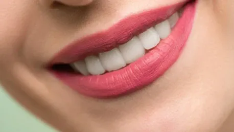
Human lips, being complex structures formed of mucosa, skin, and innervated skeletal muscles, are essential for various daily activities such as eating, drinking, talking, and social interactions. This makes their reconstruction, especially after traumatic injuries or cancer surgeries, a challenging task. Traditional surgical approaches have their limitations, often resulting in non-functional, stiff lips that do not adequately restore muscle volume or reinnervation. Advances in tissue engineering and regenerative medicine (TE/RM) promise new horizons. One promising method is the use of prelaminated flaps, specifically prelaminated innervated prevascularized prefabricated (PIPP) microvascular free flaps. This article delves into this method, presenting it as a potential breakthrough in functional lip reconstruction.
Gathering Materials
To start the process, scientists utilized previously frozen adult human keratinocytes from oral and skin tissues to fabricate a composite laminate. Human skin samples from breast reduction surgeries and oral mucosa explants from the upper inner lip were sourced from surgical discards at the University of Michigan Health System. Institutional Review Board approval was obtained for using these tissues, and donors gave their informed consent for research purposes. Preparing and obtaining the necessary materials is a crucial first step, setting the foundation for creating the mucocutaneous construct required for prelamination.
Preparing Explants
In this phase, explants were meticulously prepared, and keratinocytes were extracted by following established protocols. The protocols used, originally reported by Marcelo in 1978 for skin, and by Izumi et al. in 2000 and 2004 for oral mucosa, outlined a clear and replicable process. Specifically, tissue samples were digested with a 0.04% trypsin solution for oral tissues or a 0.125% trypsin solution for skin tissues, left to react overnight at room temperature. This enzymatic digestion facilitates the dissociation of cells from the tissue, making it possible to gather keratinocytes in a usable form.
Once the tissue samples were appropriately treated with trypsin, the dissociated keratinocytes were collected and prepared for cell culturing. Placing dissociated keratinocytes in a culture medium allows them to proliferate, providing a steady supply of these cells for further steps. Precise methodology in preparing these explants is crucial for maintaining cellular integrity and ensuring high viability rates, which are paramount for the subsequent development of a viable, functional tissue construct.
Culturing Cells
Once successfully isolated, keratinocytes were plated at a density of 7.0 · 106 cells per T-25 flask. Culturing these cells without a feeder layer, fetal bovine serum, or bovine pituitary extract was essential to maintain an environment free from animal products. For this purpose, an animal product-free culture medium, EpiLife M-EPI-500, supplemented with EpiLife Defined Growth Supplement (EDGS), was used. Additionally, the medium was enhanced with 25 mg/mL of Gentamicin and 0.375 mg/mL of Amphotericin B to support cellular growth and prevent contamination.
The primary keratinocytes proliferated in this controlled environment until reaching 70% to 80% confluence. At this stage, they were harvested and subcultured for later use in fabricating the composite laminate. Ensuring that the cells reached the correct confluence was critical for their subsequent applications, as it allowed them to expand to a sufficient number while maintaining the necessary properties for creating a functional tissue construct.
Creating Composite Laminate
In this part of the process, both oral and skin keratinocytes were expanded in a serum-free, low-calcium medium (0.06 mM CaCl2). The need for autogenous oral keratinocyte amplification, due to typically smaller oral explants, dictated the time required for this step. Once cultured and grown in separate flasks, the primary keratinocytes were frozen after reaching passage two. Thawed when needed, these cells were further cultured to achieve the requisite number for seeding onto the dermal allograft at a specified density of 2.5 x 10⁵ cells/cm2.
The next stage of the protocol necessitated presoaking AlloDerm® in 5 mg/cm2 of human type IV collagen for at least three hours before keratinocyte seeding. This step is crucial as it prepares the dermal allograft by creating an optimal environment for keratinocyte adhesion and growth. The manufacturing protocol can vary in time, particularly due to the need for amplifying oral keratinocytes, underscoring the importance of precise calculations relative to the size of the surgical grafting site.
Seeding Keratinocytes
After the growth of enough skin and oral keratinocytes in the culture flasks, the next step involved seeding these cell populations onto AlloDerm® that had been coated with type IV collagen. To create a cohesive tissue composite, keratinocytes were seeded in two separate compartments on the same AlloDerm® piece, utilizing patented custom culture ware that permitted the formation of an interface between the two keratinocyte populations. This process involves meticulously growing human oral and skin keratinocytes into a three-dimensional construct, forming a mucocutaneous (M/C) junction containing stratified oral mucosa and skin layers on the shared dermal component.
Ensuring the cells were seeded in separately compartmentalized areas was key to emulating the natural anatomy of the lips, which features distinct zones of mucosa and skin. This precise partitioning allows the creation of a 3D construct with an interface mimicking the natural transitional zone found in situ. The importance of this step cannot be overstated, as it lays the groundwork for developing a tissue composite that is structurally and functionally similar to the native lip.
Raising Construct to Air-Liquid Interface
Following the seeding of keratinocytes, the oral and skin cells were kept separate overnight by a temporary, removable barrier made of a biocompatible material like silicone. After spending several days in culture, the entire construct was exposed to an air-liquid interface, evolving into a 3D organotypic construct. This key transition forms a distinct zone between the oral and skin keratinocytes. Phenotypic markers specific to each cell type confirm the presence of three distinct areas that replicate lip epithelium as found in situ.
To aid the production of mucocutaneous (M/C) laminate, patented barrier devices were used. These tools help create a tissue construct that reflects the anatomy of human lips. This air-liquid interface step is pivotal because it promotes the differentiation and maturation of keratinocytes, resulting in a layered tissue structure that mimics the functional and aesthetic attributes of natural lips. Successfully forming this transitional zone is crucial for replicating the dynamic and composite nature of human lips, opening new avenues for reconstructive surgery.
The method described showcases the potential of cutting-edge engineered tissue constructs to transform functional human lip reconstruction. By addressing complex issues like tissue complexity and vascularity and providing innovative solutions, this hybrid approach combining in vitro fabrication with in situ muscle flap prefabrication sets a promising path for future advancements in tissue engineering and regenerative medicine (TE/RM). Developing prelaminated flaps marks a significant leap toward restoring full functionality and aesthetics for individuals with extensive lip loss.


