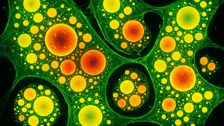
This how-to guide aims to provide a detailed roadmap for researchers, scientists, and students in cellular biology to understand and replicate the groundbreaking technique of visualizing lipid transport in living cells using fluorescence microscopy. Imagine a world where the invisible logistics of cellular life are laid bare, revealing how lipids, the essential building blocks of cell membranes, move dynamically within living systems. Historically, tracking these tiny molecules has been a monumental challenge, leaving critical questions about cellular health unanswered. With metabolic and neurodegenerative diseases linked to lipid imbalances affecting millions globally, the urgency to decode these processes has never been greater. This guide unveils a revolutionary method developed by leading researchers to observe lipid transport in real time, offering a window into cellular function that could redefine disease research. By following these steps, readers will gain insights into crafting tools and applying techniques to unlock the mysteries of lipid movement, potentially contributing to life-changing medical advancements.
Unveiling Cellular Secrets: A New Era in Lipid Imaging
The ability to visualize lipid transport within living cells marks a transformative leap in cellular biology, shedding light on processes once shrouded in mystery. Lipids are fundamental to cell structure and function, yet their rapid, intricate movements between organelles have eluded scientists due to limitations in traditional imaging methods. This guide introduces a cutting-edge fluorescence microscopy technique pioneered by teams at renowned institutions such as the Max Planck Institute of Molecular Cell Biology and Genetics (MPI-CBG) and the Biotechnology Center (BIOTEC) at TUD Dresden University of Technology. Their innovation promises to reveal the hidden choreography of cellular logistics, offering unprecedented clarity on how cells maintain balance and respond to stress.
Beyond mere observation, this breakthrough holds the potential to revolutionize the understanding of diseases tied to lipid dysregulation. Conditions like nonalcoholic fatty liver disease and neurodegenerative disorders often stem from disrupted lipid distribution, making this method a vital tool for identifying therapeutic targets. The excitement surrounding this advancement lies in its capacity to bridge the gap between basic science and clinical application, paving the way for novel treatments.
This guide is crafted to empower readers with the knowledge and practical steps to replicate and build upon this technique. By mastering the visualization of lipid transport, researchers can contribute to solving some of the most pressing health challenges of our time. The following sections detail the significance of lipids, the methodology behind the imaging process, and the broader implications of these findings, ensuring a comprehensive understanding of this scientific milestone.
Why Lipid Transport Matters: The Backbone of Cellular Health
Lipids serve as indispensable components of cellular life, forming the membranes that compartmentalize organelles, acting as energy reservoirs, and playing key roles in signaling pathways. Their proper distribution across cellular structures is critical for maintaining homeostasis, as each organelle requires a unique lipid composition to function optimally. Disruptions in this delicate balance can lead to severe health issues, including metabolic disorders and neurodegenerative conditions, highlighting the need for precise tools to study lipid dynamics.
Historically, observing lipid transport has posed significant obstacles due to the small size of these molecules and their lack of natural fluorescence, rendering them invisible to conventional microscopy. This limitation has hindered progress in understanding how cells manage lipid sorting and movement, leaving gaps in knowledge about fundamental biological processes. The inability to track lipids in real time has also constrained efforts to link specific transport mechanisms to disease states, stalling potential interventions.
The urgency to overcome these barriers cannot be overstated, as lipid imbalances are implicated in a wide array of pathologies affecting millions worldwide. By providing a method to visualize lipid transport, this guide addresses a critical need in cellular research, offering a pathway to uncover the mechanisms that underpin health and disease. Readers will learn how this innovative approach not only enhances scientific inquiry but also opens doors to therapeutic discoveries that could transform patient outcomes.
Decoding the Method: How Fluorescence Microscopy Tracks Lipids
This section outlines a meticulous, step-by-step process to visualize lipid transport in living cells using fluorescence microscopy, as developed by the Dresden research team. These instructions are designed for researchers with access to advanced laboratory equipment and a background in cellular biology, ensuring accurate replication of the technique. Each step is accompanied by detailed explanations to facilitate understanding and implementation.
Step 1: Crafting Bifunctional Lipids for Tracking
The first crucial step involves synthesizing bifunctional lipids that closely mimic natural lipids while incorporating chemical modifications for imaging purposes. These engineered molecules are designed to integrate seamlessly into cellular membranes without disrupting normal function. Researchers must use chemical synthesis techniques to attach specific tags to the lipids, enabling their detection under specialized microscopy conditions.
Precision in Design: Mimicking Natural Behavior
To ensure the bifunctional lipids behave like their natural counterparts, careful attention must be paid to their molecular structure during synthesis. The goal is to minimize alterations that could affect their integration into cell membranes or their transport pathways. This precision is vital for obtaining reliable data that reflects true lipid dynamics within living systems, avoiding artifacts that could skew results.
Chemical Tags: Enabling Visibility
The chemical tags attached to these lipids are selected for their ability to respond to ultraviolet (UV) light activation, making the lipids visible under fluorescence microscopy. These tags must be stable within the cellular environment yet reactive enough to form detectable signals when exposed to specific wavelengths. This dual functionality allows researchers to track the lipids’ locations and movements with high accuracy, a cornerstone of the imaging process.
Step 2: Loading and Observing Lipids in Living Cells
Once the bifunctional lipids are synthesized, the next step is to introduce them into living human cells for observation. Cells are typically cultured under controlled conditions, and the modified lipids are loaded into their membranes using established biochemical methods. Over a designated period, researchers monitor how these lipids distribute and integrate into various cellular structures.
Choosing the Right Cells: Ideal Candidates for Imaging
Selecting appropriate cell types is essential for successful imaging outcomes, with human bone or intestinal cells often chosen for their suitability in fluorescence studies. These cell lines offer clear visibility of membrane structures and robust lipid transport activity, making them ideal for capturing dynamic processes. The choice of cell type should align with the specific research objectives and imaging capabilities available in the laboratory.
Tracking Movement: Capturing Real-Time Dynamics
After loading the lipids, continuous observation using time-lapse microscopy allows researchers to capture their movement across organelle membranes in real time. This step requires precise calibration of imaging equipment to detect subtle changes in lipid localization over time. Documenting these dynamics provides critical insights into the pathways and mechanisms governing lipid transport within the cellular environment.
Step 3: Activating and Mapping with UV Light
The third step involves activating the chemical tags on the bifunctional lipids using UV light, which triggers a crosslinking reaction with nearby proteins. This reaction stabilizes the lipids’ positions, making them detectable under a fluorescence microscope. Researchers must carefully control the intensity and duration of UV exposure to avoid cellular damage while ensuring clear visualization.
Binding for Clarity: The Crosslinking Advantage
Crosslinking offers a significant advantage by anchoring the lipids to nearby proteins, preserving their spatial arrangement during imaging. This binding mechanism ensures that the lipids remain visible even as they traverse complex cellular networks, providing a stable signal for tracking. Such clarity is essential for mapping the intricate routes of lipid exchange between organelles with precision.
Step 4: Analyzing Data with AI and Mass Spectrometry
The final step focuses on processing and interpreting the vast datasets generated from imaging experiments. Advanced computational tools, including artificial intelligence (AI), are employed to segment images and quantify lipid flow across cellular compartments. Additionally, ultra-high-resolution mass spectrometry is used to examine structural changes in lipids during transport, adding depth to the analysis.
AI Assistance: Automating Complex Analysis
AI-driven algorithms play a pivotal role in managing the extensive imaging data, automating the segmentation of cellular structures and mapping lipid distribution patterns. These tools enhance efficiency by identifying trends and anomalies that might be missed through manual analysis. Leveraging AI ensures that the quantification of lipid transport is both accurate and reproducible, a critical factor for scientific validation.
Structural Insights: Mass Spectrometry’s Role
Complementing AI analysis, mass spectrometry provides detailed information on the molecular composition and structural transformations of lipids as they move through the cell. This technique reveals how specific lipid species adapt during transport, offering a deeper understanding of their biochemical interactions. Integrating these insights with imaging data creates a comprehensive picture of lipid behavior at the molecular level.
Key Takeaways: Redefining Lipid Transport Mechanisms
The findings from this innovative technique have reshaped the understanding of lipid transport, providing critical insights into cellular logistics. A staggering 85-95% of lipid movement occurs through non-vesicular transport mediated by carrier proteins, rather than through membrane-bound vesicles, challenging long-held assumptions. This protein-driven mechanism is not only predominant but also remarkably efficient in maintaining organelle-specific lipid profiles.
Furthermore, the speed of protein-mediated transport stands out, operating ten times faster than vesicular methods, ensuring rapid cellular responses. This specificity targets particular lipid types to distinct organelles, a process vital for cellular function. The research also delivers the first quantitative map of lipid flow across cellular compartments, a landmark achievement in visualizing these dynamics.
Finally, the selective nature of non-vesicular transport underpins cellular homeostasis by preserving unique lipid compositions for each organelle. These discoveries, distilled from meticulous imaging and analysis, highlight the precision and adaptability of cellular systems. They serve as a foundation for readers to appreciate the complexity of lipid transport and its implications for broader biological research.
Broader Impact: Transforming Disease Research and Beyond
The ability to visualize lipid transport extends far beyond academic curiosity, offering tangible benefits for medical science. By mapping lipid flow, this technique aids in pinpointing disruptions linked to diseases such as nonalcoholic fatty liver disease and neurodegenerative disorders, which affect countless individuals worldwide. Identifying specific transport pathways and proteins involved could lead to novel drug targets, accelerating the development of targeted therapies.
Moreover, the implications of this research touch on preventive medicine, as understanding lipid dynamics may inform strategies to mitigate disease risk before symptoms emerge. Challenges remain, including the need to identify exact lipid-transfer proteins and the energy sources driving transport processes. These unresolved questions present opportunities for further investigation, building on the foundation established by this methodology.
Looking toward future trends, the integration of molecular-level analysis into cellular studies is gaining momentum, fueled by interdisciplinary collaboration. Combining expertise in chemical biology, imaging technology, and computational modeling exemplifies how diverse fields can converge to tackle complex problems. This collaborative spirit ensures that advancements in lipid research will continue to evolve, potentially reshaping approaches to health and disease management on a global scale.
Looking Ahead: A Milestone and a Starting Point
Reflecting on the journey of implementing this fluorescence microscopy technique, the steps taken to visualize lipid transport in living cells marked a turning point in cellular biology. The meticulous synthesis of bifunctional lipids, their integration into living systems, the activation via UV light, and the sophisticated analysis using AI and mass spectrometry all converged to reveal the dominance of non-vesicular, protein-mediated transport. These efforts provided a clearer picture of how cells orchestrate lipid distribution with speed and precision.
As a next step, researchers are encouraged to delve deeper into identifying specific lipid-transfer proteins and exploring the energy mechanisms behind transport processes. Collaborating across disciplines and leveraging emerging technologies can further refine this technique, addressing remaining gaps in knowledge. Engaging with ongoing studies and contributing to databases of lipid dynamics will amplify the impact of these findings.
Additionally, applying this method to diverse cell types and disease models could uncover tailored insights, guiding the development of personalized treatments. The path forward involves not only building on the quantitative map of lipid flow but also translating these discoveries into clinical solutions. This milestone, while significant, serves as a springboard for continued exploration, urging the scientific community to push boundaries in understanding cellular intricacies.
