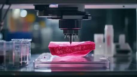

Bioprinting has emerged as a revolutionary bioengineering technique that utilizes the principles of 3D printing to create tissue-like structures embedded with living cells. Since its inception in 1988, this technology has experienced tremendous growth, advancing from its original concept to today’s more sophisticated applications that span tissue engineering, drug discovery, and more. The developments in bioprinting have not only enhanced the capabilities of creating synthetic biological structures but have also opened new avenues for medical research and clinical applications.
Breakthroughs in High-Density Tissue Structures
One of the most significant advancements in bioprinting involves the creation of high-density tissue structures, overcoming a major challenge in scaling up tissue production. Researchers at Penn State University have developed a technique called the High-throughput Integrated Tissue Fabrication System for Bioprinting (HITS-Bio) to address this issue. By using spheroids—dense clusters of cells that closely mimic the cell density of the human body—HITS-Bio employs a digitally controlled array of multiple nozzles that move in three dimensions to effectively manipulate these spheroids. This innovative approach allows for bioprinting at a speed ten times faster than previous methods while maintaining cell viability above 90%.
The benefits of this breakthrough are profound, particularly for applications requiring high-density tissues. For example, the successful bioprinting of cartilage tissue using the HITS-Bio technique showcases its potential for repairing damaged tissue. Furthermore, experiments in rat models have demonstrated that this method is effective in wound repair, indicating practical applications in regenerative medicine. The success of this technique not only marks a significant leap in the speed and efficiency of bioprinting but also paves the way for more complex tissue engineering projects in the future.
Innovations in Neurodegenerative Disease Research
The scope of bioprinting extends far beyond traditional tissue engineering, penetrating areas like neurodegenerative disease research. Pioneering work by Marimélia Porcionatto and her team has focused on biofabricating 3D systems to study the central nervous system. The creation of these 3D models represents a significant advancement in the intersection of bioprinting and neurological research. Though details on specific methodologies were not provided, this initiative marks an exciting development, particularly in studying conditions such as Alzheimer’s disease.
The ability to replicate the complex environment of the central nervous system using 3D bioprinting offers unprecedented opportunities for understanding the mechanisms behind neurodegenerative diseases. These biomimetic models could drastically improve the way researchers investigate disease progression and test potential treatments, ultimately contributing to the development of more effective therapeutic strategies. The integration of bioprinting technology in neuro research underscores its potential to transform our approach to complex medical challenges.
Shape-Morphing Heart Tissues
Another landmark in bioprinting innovations is the development of shape-morphing human heart tissues. At the University of Galway, researchers led by Ankita Pramanick and Andrew Daly are working on creating heart tissues that can change shape dynamically, thereby enhancing their contractile strength. This research highlights the importance of cell-shape changes during embryonic development for the formation of mature heart tissue. Using a novel platform that enables embedded bioprinting, the team has designed tissues capable of programmable, predictable 4D shape changes driven by cell-generated forces.
By modifying print geometry and adjusting the stiffness of the bioink, the researchers have succeeded in producing heart tissues that closely mimic the contraction strength of human adult heart tissue. This advancement holds significant implications for heart tissue repair and simulation, offering new hope for patients with cardiac conditions. The ability to replicate the natural dynamics of heart tissue more accurately points to a future where bioprinted tissues could be used in clinical settings to repair damaged hearts or even replace failing heart tissue.
Tumor Microenvironments and Drug Response
The application of bioprinting in oncology is another promising frontier. By creating accurate tumor microenvironments using 3D bioprinting technology, researchers can now develop cancer models that retain the original characteristics of tumors. This capability allows for more realistic assessments of drug responses, potentially leading to the discovery of more effective cancer therapies. The precision and reproducibility offered by bioprinting ensure that these models can be used to systematically evaluate the efficacy of various treatments under conditions that closely mimic human physiology.
Current developments in this field have proved instrumental in advancing our understanding of tumor biology. By accurately replicating the intricate details of tumor environments, bioprinting has enabled researchers to observe how cancer cells interact with their surrounding tissues and respond to different drugs. This enhanced understanding can bring about significant improvements in the design of preclinical trials, ultimately leading to better-targeted therapies and personalized medicine approaches for cancer treatment. The continued integration of bioprinting technology in oncology research promises to revolutionize cancer diagnostics and therapy.
Blood-Brain Barrier Models
Studying the blood-brain barrier (BBB) is crucial for understanding several neurodegenerative diseases, and bioprinting has made significant contributions in this area as well. Scientists at Pohang University of Science and Technology (POSTECH) and Seoul National University Hospital have developed a 3D bioprinted model to study the BBB using cerebrovascular-specific bioinks derived from decellularized extracellular matrix (CBVdECM) from pig brains and blood vessels. This bioink, combined with 3D bioprinting technology, allows for the creation of tubular vascular models that accurately replicate the structure and function of the BBB.
These bioprinted models respond appropriately to inflammatory substances, providing crucial insights into BBB dysfunction and inflammation associated with neurodegenerative diseases like Alzheimer’s. The ability to mimic the human BBB’s complex structure and response enhances our understanding of how this barrier operates under pathological conditions. Researchers aim to further refine these models by integrating additional cell types and improving methods to quantify inflammatory responses and barrier permeability, thereby expanding their application in patient-specific disease models. This research represents a significant step forward in our ability to study and potentially treat neurodegenerative diseases.
Sustainable and Innovative Bioinks
The innovation in bioprinting is not just limited to creating 3D biological structures; it also includes the development of sustainable and efficient bioinks. BIO INX, a company based in Belgium, has introduced BIORES INX, a gelatin methacrylamide-based resin designed for light-based bioprinting. Complying with biocompatibility standards such as ISO 10993-5, this bioink offers simplicity in processing and compatibility with various digital light-processing bioprinting platforms. Its introduction marks a significant step forward in creating more reliable and versatile bioinks for complex tissue engineering tasks.
In addition, researchers at Seoul National University of Science and Technology have developed a sustainable nanocellulose bioink derived from Kombucha SCOBY. By reinforcing this naturally produced cellulose with chitosan and kaolin nanoparticles, they created a stable hydrogel suitable for 3D printing. This bioink can be loaded into a handheld ‘Biowork’ biopen along with living cells, allowing for precise application to damaged tissue areas. The sustainable nature and practical applications of this bioink demonstrate its potential for immediate and effective wound treatment, presenting a viable alternative to traditional materials used in bioprinting.
Approaches to Wound Healing
Innovative approaches to wound healing have also been explored using the potential of bioprinting. One notable example involves the use of bioactive hydrogel ink containing macrophage-derived extracellular vesicles, which enhance the healing of cutaneous wounds. Delivered in situ using a 3D-printing pen, this method significantly advances therapeutic strategies for wound treatment and tissue repair. The hydrogel ink not only promotes faster wound healing but also represents a new frontier in bioengineering by integrating biological components that enhance the body’s natural healing processes.
The clinical applications of this technology are far-reaching, offering new solutions for patients with chronic wounds or those recovering from surgical procedures. The ability to precisely deliver bioactive compounds to the site of injury using bioprinted materials underscores the transformative potential of bioprinting in medical treatments. As research in this area continues, we can expect to see further advancements that will improve the efficacy of wound healing protocols and expand the use of bioprinting in various therapeutic contexts.
Bone and Soft Tissue Repair
Advancements in bioprinting have also led to significant progress in bone and soft tissue repair. Researchers at Hefei Institutes of Physical Science, part of the Chinese Academy of Sciences, have developed innovative 3D bioprinting materials that combine boron-based glass (BBG) with polycaprolactone (PCL) and sodium alginate. These composites have been optimized for bone healing, with research indicating that a BBG/PCL composite containing 20% BBG is ideal for bone regeneration. Meanwhile, their BBG/sodium alginate bioink has shown high precision, structural integrity, and biocompatibility, making it suitable for soft tissue repair.
This research is particularly significant because it addresses the challenge of creating materials that are both strong enough to support bone regeneration and flexible enough to facilitate soft tissue repair. The success of these bioprinted materials highlights the potential for developing more effective treatment options for patients with injuries to bones and soft tissues. By leveraging the unique properties of these composites, bioprinting can offer new solutions for complex medical conditions, further demonstrating its versatility and transformative impact on the field of regenerative medicine.
Expanding Horizons in Bioprinting
Bioprinting, a groundbreaking bioengineering technique that applies 3D printing principles to produce tissue-like structures with living cells, has revolutionized the field since its inception in 1988. This technology has seen tremendous growth, evolving from a novel idea to advanced applications in tissue engineering, drug discovery, and more. The advances in bioprinting have significantly improved the ability to create synthetic biological structures and have paved the way for new possibilities in medical research and clinical applications. Bioprinting now plays a crucial role in developing complex tissue models that mimic human organs and tissues, offering a valuable tool for understanding diseases, testing new drugs, and potentially creating functional organs for transplantation. Furthermore, the ongoing innovations in bioprinting techniques and materials are setting the stage for even more groundbreaking advancements in the future. This technology is poised to make substantial contributions to regenerative medicine, personalized healthcare, and beyond.